6TSI | pdb_00006tsi

cd1 nitrite reductase NirS with bound dihydro-heme d1


Experimental Data Snapshot

  • Method: X-RAY DIFFRACTION
  • Resolution: 2.38 Å
  • R-Value Free: 
    0.280 (Depositor), 0.257 (DCC) 
  • R-Value Work: 
    0.238 (Depositor) 
  • R-Value Observed: 
    0.240 (Depositor) 

Starting Model: experimental
View more details

wwPDB Validation 3D Report Full Report

Validation slider image for 6TSI

Ligand Structure Quality Assessment 


This is version 1.2 of the entry. See complete history

Literature

Structure of heme d 1 -free cd 1 nitrite reductase NirS.

Klunemann, T.Blankenfeldt, W.

(2020) Acta Crystallogr F Struct Biol Commun 76: 250-256

  • DOI: https://doi.org/10.1107/S2053230X20006676
  • Primary Citation Related Structures: 
    6TPO, 6TSI

  • PubMed Abstract: 

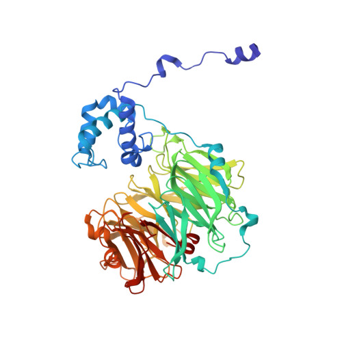
    A key step in anaerobic nitrate respiration is the reduction of nitrite to nitric oxide, which is catalysed by the cd 1 nitrite reductase NirS in, for example, the Gram-negative opportunistic pathogen Pseudomonas aeruginosa. Each subunit of this homodimeric enzyme consists of a cytochrome c domain and an eight-bladed β-propeller that binds the uncommon isobacteriochlorin heme d 1 as an essential part of its active site. Although NirS has been well studied mechanistically and structurally, the focus of previous studies has been on the active heme d 1 -bound form. The heme d 1 -free form of NirS reported here, which represents a premature state of the reductase, adopts an open conformation with the cytochrome c domains moved away from each other with respect to the active enzyme. Further, the movement of a loop around Trp498 seems to be related to a widening of the propeller, allowing easier access to the heme d 1 -binding side. Finally, a possible link between the open conformation of NirS and flagella formation in P. aeruginosa is discussed.


  • Organizational Affiliation
    • Structure and Function of Proteins, Helmholtz Centre for Infection Research, Inhoffenstrasse 7, 38124 Braunschweig, Niedersachsen, Germany.

Macromolecule Content 

  • Total Structure Weight: 123.83 kDa 
  • Atom Count: 8,824 
  • Modeled Residue Count: 1,080 
  • Deposited Residue Count: 1,086 
  • Unique protein chains: 1

Macromolecules

Find similar proteins by:|  3D Structure
Entity ID: 1
MoleculeChains  Sequence LengthOrganismDetailsImage
Nitrite reductase
A, B
543Pseudomonas aeruginosa PAO1Mutation(s): 0 
EC: 1.7.2.1 (PDB Primary Data), 1.7.99.1 (PDB Primary Data)
UniProt
Find proteins for P24474 (Pseudomonas aeruginosa (strain ATCC 15692 / DSM 22644 / CIP 104116 / JCM 14847 / LMG 12228 / 1C / PRS 101 / PAO1))
Explore P24474 
Go to UniProtKB:  P24474
Entity Groups
Sequence Clusters30% Identity50% Identity70% Identity90% Identity95% Identity100% Identity
UniProt GroupP24474
Sequence Annotations
Expand
Reference Sequence

Small Molecules

Ligands 4 Unique
IDChains Name / Formula / InChI Key2D Diagram3D Interactions
DHE
(Subject of Investigation/LOI)

Query on DHE


Download:Ideal Coordinates CCD File
D [auth A],
K [auth B]
HEME D
C34 H32 Fe N4 O10
XLQCGNUTSJTZNF-YDXXJHAFSA-L
HEC

Query on HEC


Download:Ideal Coordinates CCD File
C [auth A],
J [auth B]
HEME C
C34 H34 Fe N4 O4
HXQIYSLZKNYNMH-LJNAALQVSA-N
PO4

Query on PO4


Download:Ideal Coordinates CCD File
E [auth A],
F [auth A],
G [auth A],
L [auth B],
M [auth B]
PHOSPHATE ION
O4 P
NBIIXXVUZAFLBC-UHFFFAOYSA-K
CL

Query on CL


Download:Ideal Coordinates CCD File
H [auth A],
I [auth A],
N [auth B],
O [auth B],
P [auth B]
CHLORIDE ION
Cl
VEXZGXHMUGYJMC-UHFFFAOYSA-M

Experimental Data & Validation

Experimental Data

  • Method: X-RAY DIFFRACTION
  • Resolution: 2.38 Å
  • R-Value Free:  0.280 (Depositor), 0.257 (DCC) 
  • R-Value Work:  0.238 (Depositor) 
  • R-Value Observed: 0.240 (Depositor) 
Space Group: P 21 21 2
Unit Cell:
Length ( Å )Angle ( ˚ )
a = 163.762α = 90
b = 91.093β = 90
c = 112.706γ = 90
Software Package:
Software NamePurpose
PHENIXrefinement
XDSdata reduction
autoPROCdata collection
STARANISOdata scaling
PDB_EXTRACTdata extraction
PHENIXphasing

Structure Validation

View Full Validation Report


Ligand Structure Quality Assessment 


Entry History 

& Funding Information

Deposition Data


Funding OrganizationLocationGrant Number
German Research Foundation (DFG)GermanyGRK2223/1

Revision History  (Full details and data files)

  • Version 1.0: 2020-11-18
    Type: Initial release
  • Version 1.1: 2024-01-24
    Changes: Data collection, Database references, Refinement description
  • Version 1.2: 2024-11-13
    Changes: Structure summary