6TRX | pdb_00006trx

Crystal structure of DPP8 in complex with 1G244


Experimental Data Snapshot

  • Method: X-RAY DIFFRACTION
  • Resolution: 3.20 Å
  • R-Value Free: 
    0.238 (Depositor), 0.236 (DCC) 
  • R-Value Work: 
    0.197 (Depositor), 0.200 (DCC) 
  • R-Value Observed: 
    0.199 (Depositor) 

Starting Model: experimental
View more details

wwPDB Validation 3D Report Full Report

Validation slider image for 6TRX

Ligand Structure Quality Assessment 


This is version 1.3 of the entry. See complete history

Literature

Aerosol-based ligand soaking of reservoir-free protein crystals.

Ross, B.Krapp, S.Geiss-Friedlander, R.Littmann, W.Huber, R.Kiefersauer, R.

(2021) J Appl Crystallogr 54: 895-902

  • DOI: https://doi.org/10.1107/S1600576721003551
  • Primary Citation Related Structures: 
    6TRW, 6TRX, 7NVQ

  • PubMed Abstract: 

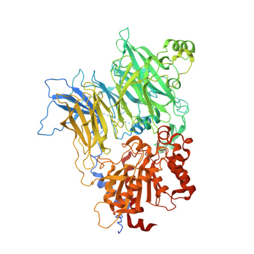
    Soaking of macromolecular crystals allows the formation of complexes via diffusion of molecules into a preformed crystal for structural analysis. Soaking offers various advantages over co-crystallization, e.g. small samples and high-throughput experimentation. However, this method has disadvantages, such as inducing mechanical stress on crystals and reduced success rate caused by low affinity/solubility of the ligand. To bypass these issues, the Picodropper was previously developed in the authors' laboratory. This technique aimed to deliver small volumes of compound solution in response to crystal dehydration supported by the Free Mounting System humidity control or by IR-laser-induced protein crystal transformation. Herein, a new related soaking development, the Aerosol-Generator, is introduced. This device delivers compounds onto the solution-free surface of protein crystals using an ultrasonic technique. The produced aerosol stream enables an easier and more accurate control of solution volumes, reduced crystal handling, and crystal-size-independent soaking. The Aerosol-Generator has been used to produce complexes of DPP8 crystals, where otherwise regular soaking did not achieve complex formation. These results demonstrate the potential of this device in challenging ligand-binding scenarios and contribute to further understanding of DPP8 inhibitor binding.


  • Organizational Affiliation
    • Max Planck Institut für Biochemie, D-82152 Martinsried, Germany.

Macromolecule Content 

  • Total Structure Weight: 313.75 kDa 
  • Atom Count: 20,800 
  • Modeled Residue Count: 2,523 
  • Deposited Residue Count: 2,709 
  • Unique protein chains: 1

Macromolecules

Find similar proteins by:|  3D Structure
Entity ID: 1
MoleculeChains  Sequence LengthOrganismDetailsImage
Dipeptidyl peptidase 8A [auth B],
B [auth A],
C
903Homo sapiensMutation(s): 0 
Gene Names: DPP8DPRP1MSTP097MSTP135MSTP141
EC: 3.4.14.5
UniProt & NIH Common Fund Data Resources
Find proteins for Q6V1X1 (Homo sapiens)
Explore Q6V1X1 
Go to UniProtKB:  Q6V1X1
PHAROS:  Q6V1X1
GTEx:  ENSG00000074603 
Entity Groups
Sequence Clusters30% Identity50% Identity70% Identity90% Identity95% Identity100% Identity
UniProt GroupQ6V1X1
Sequence Annotations
Expand
Reference Sequence

Small Molecules

Ligands 4 Unique
IDChains Name / Formula / InChI Key2D Diagram3D Interactions
9XH

Query on 9XH


Download:Ideal Coordinates CCD File
E [auth B],
I [auth A],
M [auth C]
(2~{S})-2-azanyl-4-[4-[bis(4-fluorophenyl)methyl]piperazin-1-yl]-1-(1,3-dihydroisoindol-2-yl)butane-1,4-dione
C29 H30 F2 N4 O2
ZKIQFLSGMMYCGS-SANMLTNESA-N
PO4

Query on PO4


Download:Ideal Coordinates CCD File
G [auth B],
K [auth A],
O [auth C]
PHOSPHATE ION
O4 P
NBIIXXVUZAFLBC-UHFFFAOYSA-K
TMO

Query on TMO


Download:Ideal Coordinates CCD File
D [auth B],
H [auth A],
L [auth C]
trimethylamine oxide
C3 H9 N O
UYPYRKYUKCHHIB-UHFFFAOYSA-N
NA

Query on NA


Download:Ideal Coordinates CCD File
F [auth B],
J [auth A],
N [auth C]
SODIUM ION
Na
FKNQFGJONOIPTF-UHFFFAOYSA-N
Binding Affinity Annotations 
IDSourceBinding Affinity
9XH BindingDB:  6TRX IC50: min: 12, max: 1400 (nM) from 4 assay(s)

Experimental Data & Validation

Experimental Data

  • Method: X-RAY DIFFRACTION
  • Resolution: 3.20 Å
  • R-Value Free:  0.238 (Depositor), 0.236 (DCC) 
  • R-Value Work:  0.197 (Depositor), 0.200 (DCC) 
  • R-Value Observed: 0.199 (Depositor) 
Space Group: C 2 2 21
Unit Cell:
Length ( Å )Angle ( ˚ )
a = 163.963α = 90
b = 246.288β = 90
c = 261.805γ = 90
Software Package:
Software NamePurpose
XSCALEdata scaling
REFMACrefinement
PDB_EXTRACTdata extraction
XDSdata reduction
PHASERphasing

Structure Validation

View Full Validation Report


Ligand Structure Quality Assessment 


Entry History 

Deposition Data

Revision History  (Full details and data files)

  • Version 1.0: 2021-01-13
    Type: Initial release
  • Version 1.1: 2021-06-16
    Changes: Database references
  • Version 1.2: 2021-07-14
    Changes: Database references
  • Version 1.3: 2024-01-24
    Changes: Data collection, Database references, Refinement description