A New Docking Domain Type in the Peptide-Antimicrobial-Xenorhabdus Peptide Producing Nonribosomal Peptide Synthetase fromXenorhabdus bovienii.
Watzel, J., Hacker, C., Duchardt-Ferner, E., Bode, H.B., Wohnert, J.(2020) ACS Chem Biol 15: 982-989
- PubMed: 32167274 Search on PubMed
- DOI: https://doi.org/10.1021/acschembio.9b01022
- Primary Citation Related Structures:
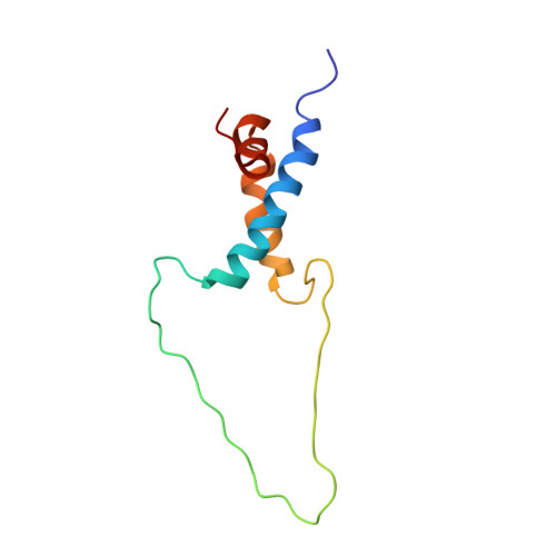
6TRP - PubMed Abstract:
Nonribosomal peptide synthetases (NRPSs) produce a wide variety of different natural products from amino acid precursors. In contrast to single protein NRPS, the NRPS of the bacterium Xenorhabdus bovienii producing the peptide-antimicrobial-Xenorhabdus (PAX) peptide consists of three individual proteins (PaxA/B/C), which interact with each other noncovalently in a linear fashion. The specific interactions between the three different proteins in this NRPS system are mediated by short C- and N-terminal docking domains ( C/N DDs). Here, we investigate the structural basis for the specific interaction between the C DD from the protein PaxB and the N DD from PaxC. The isolated DD peptides feature transient α-helical conformations in the absence of the respective DD partner. Isothermal titration calorimetry (ITC) and nuclear magnetic resonance (NMR) titration experiments showed that the two isolated DDs bind to each other and form a structurally well-defined complex with a dissociation constant in the micromolar range as is typical for many DD interactions. Artificial linking of this DD pair via a flexible glycine-serine (GS) linker enabled us to solve the structure of the DD complex by NMR spectroscopy. In the complex, the two DDs interact with each other by forming a three helix bundle arranged in an overall coiled-coil motif. Key interacting residues were identified in mutagenesis experiments. Overall, our structure of the PaxB C DD/PaxC N DD complex represents an architecturally new type of DD interaction motif.
- Molecular Biotechnology, Institute of Molecular Biosciences, Goethe University Frankfurt, 60438 Frankfurt am Main, Germany.
Organizational Affiliation: