6TQ5 | pdb_00006tq5

Alcohol dehydrogenase from Candida magnoliae DSMZ 70638 (ADHA): complex with NADP+


Experimental Data Snapshot

  • Method: X-RAY DIFFRACTION
  • Resolution: 1.60 Å
  • R-Value Free: 
    0.197 (Depositor), 0.221 (DCC) 
  • R-Value Work: 
    0.141 (Depositor) 
  • R-Value Observed: 
    0.144 (Depositor) 

Starting Model: experimental
View more details

wwPDB Validation 3D Report Full Report

Validation slider image for 6TQ5

Ligand Structure Quality Assessment 


This is version 1.1 of the entry. See complete history

Literature

Approaching boiling point stability of an alcohol dehydrogenase through computationally-guided enzyme engineering.

Aalbers, F.S.Furst, M.J.Rovida, S.Trajkovic, M.Gomez Castellanos, J.R.Bartsch, S.Vogel, A.Mattevi, A.Fraaije, M.W.

(2020) Elife 9

  • DOI: https://doi.org/10.7554/eLife.54639
  • Primary Citation Related Structures: 
    6TQ3, 6TQ5, 6TQ8

  • PubMed Abstract: 

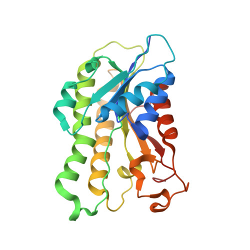
    Enzyme instability is an important limitation for the investigation and application of enzymes. Therefore, methods to rapidly and effectively improve enzyme stability are highly appealing. In this study we applied a computational method (FRESCO) to guide the engineering of an alcohol dehydrogenase. Of the 177 selected mutations, 25 mutations brought about a significant increase in apparent melting temperature (Δ T m ≥ +3 °C). By combining mutations, a 10-fold mutant was generated with a T m of 94 °C (+51 °C relative to wild type), almost reaching water's boiling point, and the highest increase with FRESCO to date. The 10-fold mutant's structure was elucidated, which enabled the identification of an activity-impairing mutation. After reverting this mutation, the enzyme showed no loss in activity compared to wild type, while displaying a T m of 88 °C (+45 °C relative to wild type). This work demonstrates the value of enzyme stabilization through computational library design.


  • Organizational Affiliation
    • Molecular Enzymology Group, University of Groningen, Groningen, Netherlands.

Macromolecule Content 

  • Total Structure Weight: 105.97 kDa 
  • Atom Count: 7,848 
  • Modeled Residue Count: 960 
  • Deposited Residue Count: 984 
  • Unique protein chains: 1

Macromolecules

Find similar proteins by:|  3D Structure
Entity ID: 1
MoleculeChains  Sequence LengthOrganismDetailsImage
Enzyme subunit
A, B, C, D
246Starmerella magnoliaeMutation(s): 0 

Small Molecules

Ligands 3 Unique
IDChains Name / Formula / InChI Key2D Diagram3D Interactions
NAP
(Subject of Investigation/LOI)

Query on NAP


Download:Ideal Coordinates CCD File
E [auth A],
G [auth B],
H [auth C],
K [auth D]
NADP NICOTINAMIDE-ADENINE-DINUCLEOTIDE PHOSPHATE
C21 H28 N7 O17 P3
XJLXINKUBYWONI-NNYOXOHSSA-N
GOL

Query on GOL


Download:Ideal Coordinates CCD File
J [auth C],
L [auth D]
GLYCEROL
C3 H8 O3
PEDCQBHIVMGVHV-UHFFFAOYSA-N
IPA

Query on IPA


Download:Ideal Coordinates CCD File
F [auth A],
I [auth C]
ISOPROPYL ALCOHOL
C3 H8 O
KFZMGEQAYNKOFK-UHFFFAOYSA-N

Experimental Data & Validation

Experimental Data

  • Method: X-RAY DIFFRACTION
  • Resolution: 1.60 Å
  • R-Value Free:  0.197 (Depositor), 0.221 (DCC) 
  • R-Value Work:  0.141 (Depositor) 
  • R-Value Observed: 0.144 (Depositor) 
Space Group: I 1 2 1
Unit Cell:
Length ( Å )Angle ( ˚ )
a = 76.115α = 90
b = 113.136β = 94.04
c = 124.206γ = 90
Software Package:
Software NamePurpose
XDSdata reduction
Aimlessdata scaling
REFMACrefinement
PDB_EXTRACTdata extraction
PHASERphasing

Structure Validation

View Full Validation Report


Ligand Structure Quality Assessment 


Entry History 

& Funding Information

Deposition Data


Funding OrganizationLocationGrant Number
European CommissionEuropean UnionH2020-LEIT BIO-2014-1 635734

Revision History  (Full details and data files)

  • Version 1.0: 2020-04-08
    Type: Initial release
  • Version 1.1: 2024-01-24
    Changes: Data collection, Database references, Refinement description