6TOP | pdb_00006top

Structure of the PorE C-terminal domain, a protein of T9SS from Porphyromonas gingivalis


Experimental Data Snapshot

  • Method: X-RAY DIFFRACTION
  • Resolution: 1.55 Å
  • R-Value Free: 
    0.215 (Depositor), 0.214 (DCC) 
  • R-Value Work: 
    0.190 (Depositor), 0.189 (DCC) 
  • R-Value Observed: 
    0.191 (Depositor) 

wwPDB Validation 3D Report Full Report

Validation slider image for 6TOP

Ligand Structure Quality Assessment 


This is version 1.1 of the entry. See complete history

Literature

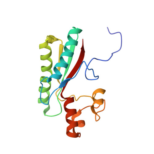
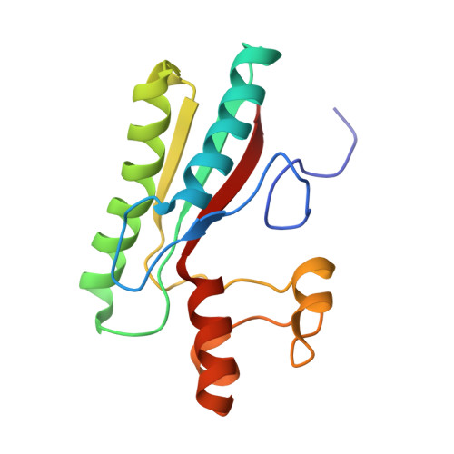
Crystal structure of Type IX secretion system PorE C-terminal domain from Porphyromonas gingivalis in complex with a peptidoglycan fragment.

Trinh, N.T.T.Tran, H.Q.Van Dong, Q.Cambillau, C.Roussel, A.Leone, P.

(2020) Sci Rep 10: 7384-7384

  • DOI: https://doi.org/10.1038/s41598-020-64115-z
  • Primary Citation Related Structures: 
    6TOP

  • PubMed Abstract: 

    Porphyromonas gingivalis, the major human pathogen associated to periodontal diseases, utilizes the Bacteroidetes-specific type IX secretion system (T9SS) to export virulence factors. PorE is a periplasmic multi-domain lipoprotein associated to the outer membrane that was recently identified as essential for T9SS function. Little is known on T9SS at the structural level, and in particular its interaction with peptidoglycan. This prompted us to carry out structural studies on PorE full length as well as on its four isolated domains. Here we report the crystal structure of the C-terminal OmpA_C-like putative peptidoglycan-binding domain at 1.55 Å resolution. An electron density volume was identified in the protein cleft, making it possible to build a naturally-occurring peptidoglycan fragment. This result suggests that PorE interacts with peptidoglycan and that PorE could anchor T9SS to the cell wall.


  • Organizational Affiliation
    • Architecture et Fonction des Macromolécules Biologiques, Aix-Marseille Université, UMR 7257, 163 Avenue de Luminy, Case 932, 13009, Marseille, France.

Macromolecule Content 

  • Total Structure Weight: 72.53 kDa 
  • Atom Count: 5,704 
  • Modeled Residue Count: 604 
  • Deposited Residue Count: 604 
  • Unique protein chains: 2

Macromolecules

Find similar proteins by:|  3D Structure
Entity ID: 1
MoleculeChains  Sequence LengthOrganismDetailsImage
OmpA family protein154Porphyromonas gingivalis JCVI SC001Mutation(s): 0 
Gene Names: A343_1565
Find similar proteins by:|  3D Structure
Entity ID: 2
MoleculeChains  Sequence LengthOrganismDetailsImage
OmpA family protein
B, C, D
150Porphyromonas gingivalis JCVI SC001Mutation(s): 0 
Gene Names: A343_1565

Small Molecules

Ligands 2 Unique
IDChains Name / Formula / InChI Key2D Diagram3D Interactions
MLD
(Subject of Investigation/LOI)

Query on MLD


Download:Ideal Coordinates CCD File
E [auth A],
G [auth B],
I [auth C],
K [auth D]
GLCNAC(BETA1-4)-MURNAC(1,6-ANHYDRO)-L-ALA-GAMMA-D-GLU-MESO-A2PM-D-ALA
C37 H59 N7 O20
UPFMKPIBAIPLHT-RSJSDIDPSA-N
NA

Query on NA


Download:Ideal Coordinates CCD File
F [auth A],
H [auth B],
J [auth C],
L [auth D]
SODIUM ION
Na
FKNQFGJONOIPTF-UHFFFAOYSA-N

Experimental Data & Validation

Experimental Data

  • Method: X-RAY DIFFRACTION
  • Resolution: 1.55 Å
  • R-Value Free:  0.215 (Depositor), 0.214 (DCC) 
  • R-Value Work:  0.190 (Depositor), 0.189 (DCC) 
  • R-Value Observed: 0.191 (Depositor) 
Space Group: C 1 2 1
Unit Cell:
Length ( Å )Angle ( ˚ )
a = 208.03α = 90
b = 52.22β = 108.38
c = 66.56γ = 90
Software Package:
Software NamePurpose
BUSTERrefinement
XDSdata reduction
XSCALEdata scaling
PHASERphasing

Structure Validation

View Full Validation Report


Ligand Structure Quality Assessment 


Entry History 

& Funding Information

Deposition Data


Funding OrganizationLocationGrant Number
French National Research AgencyFranceANR-15-CE11-0019-01

Revision History  (Full details and data files)

  • Version 1.0: 2020-06-17
    Type: Initial release
  • Version 1.1: 2024-05-15
    Changes: Data collection, Database references, Derived calculations