6THE | pdb_00006the

Crystal structure of core domain of four-domain heme-cupredoxin-Cu nitrite reductase from Bradyrhizobium sp. ORS 375


Experimental Data Snapshot

  • Method: X-RAY DIFFRACTION
  • Resolution: 2.87 Å
  • R-Value Free: 
    0.234 (Depositor), 0.234 (DCC) 
  • R-Value Work: 
    0.225 (Depositor), 0.225 (DCC) 
  • R-Value Observed: 
    0.226 (Depositor) 

Starting Model: experimental
View more details

wwPDB Validation 3D Report Full Report

Validation slider image for 6THE

This is version 1.2 of the entry. See complete history

Literature

Reverse protein engineering of a novel 4-domain copper nitrite reductase reveals functional regulation by protein-protein interaction.

Sasaki, D.Watanabe, T.F.Eady, R.R.Garratt, R.C.Antonyuk, S.V.Hasnain, S.S.

(2021) FEBS J 288: 262-280

  • DOI: https://doi.org/10.1111/febs.15324
  • Primary Citation Related Structures: 
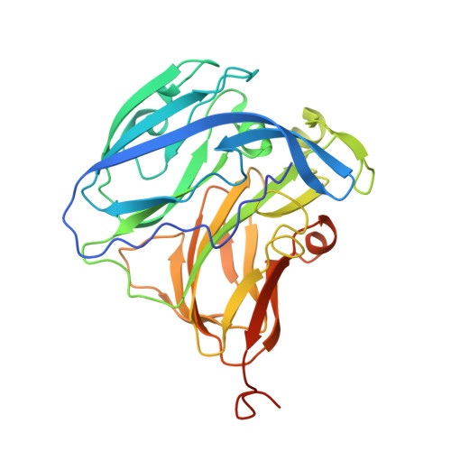
    6THE, 6THF

  • PubMed Abstract: 

    Cu-containing nitrite reductases that convert NO 2 - to NO are critical enzymes in nitrogen-based energy metabolism. Among organisms in the order Rhizobiales, we have identified two copies of nirK, one encoding a new class of 4-domain CuNiR that has both cytochrome and cupredoxin domains fused at the N terminus and the other, a classical 2-domain CuNiR (Br 2D NiR). We report the first enzymatic studies of a novel 4-domain CuNiR from Bradyrhizobium sp. ORS 375 (BrNiR), its genetically engineered 3- and 2-domain variants, and Br 2D NiR revealing up to ~ 500-fold difference in catalytic efficiency in comparison with classical 2-domain CuNiRs. Contrary to the expectation that tethering would enhance electron delivery by restricting the conformational search by having a self-contained donor-acceptor system, we demonstrate that 4-domain BrNiR utilizes N-terminal tethering for downregulating enzymatic activity instead. Both Br 2D NiR and an engineered 2-domain variant of BrNiR (Δ(Cytc-Cup) BrNiR) have 3 to 5% NiR activity compared to the well-characterized 2-domain CuNiRs from Alcaligenes xylosoxidans (AxNiR) and Achromobacter cycloclastes (AcNiR). Structural comparison of Δ(Cytc-Cup) BrNiR and Br 2D NiR with classical 2-domain AxNiR and AcNiR reveals structural differences of the proton transfer pathway that could be responsible for the lowering of activity. Our study provides insights into unique structural and functional characteristics of naturally occurring 4-domain CuNiR and its engineered 3- and 2-domain variants. The reverse protein engineering approach utilized here has shed light onto the broader question of the evolution of transient encounter complexes and tethered electron transfer complexes. ENZYME: Copper-containing nitrite reductase (CuNiR) (EC 1.7.2.1). DATABASE: The atomic coordinate and structure factor of Δ(Cytc-Cup) BrNiR and Br 2D NiR have been deposited in the Protein Data Bank (http://www.rcsb.org/) under the accession code 6THE and 6THF, respectively.


  • Organizational Affiliation
    • Molecular Biophysics Group, Institute of Systems, Molecular and Integrative Biology, Faculty of Health and Life Sciences, University of Liverpool, UK.

Macromolecule Content 

  • Total Structure Weight: 36.62 kDa 
  • Atom Count: 2,367 
  • Modeled Residue Count: 299 
  • Deposited Residue Count: 321 
  • Unique protein chains: 1

Macromolecules

Find similar proteins by:|  3D Structure
Entity ID: 1
MoleculeChains  Sequence LengthOrganismDetailsImage
Copper-containing nitrite reductase321Bradyrhizobium sp. ORS 375Mutation(s): 0 
Gene Names: BRAO375_2740002
EC: 1.7.2.1
UniProt
Find proteins for A0ACD6B9I4 (Bradyrhizobium sp. (strain ORS 375))
Explore A0ACD6B9I4 
Go to UniProtKB:  A0ACD6B9I4
Entity Groups
Sequence Clusters30% Identity50% Identity70% Identity90% Identity95% Identity100% Identity
UniProt GroupA0ACD6B9I4
Sequence Annotations
Expand
Reference Sequence

Small Molecules

Ligands 7 Unique
IDChains Name / Formula / InChI Key2D Diagram3D Interactions
YB

Query on YB


Download:Ideal Coordinates CCD File
D [auth A],
E [auth A],
F [auth A]
YTTERBIUM (III) ION
Yb
AWSFICBXMUKWSK-UHFFFAOYSA-N
ER3

Query on ER3


Download:Ideal Coordinates CCD File
G [auth A],
H [auth A],
I [auth A],
J [auth A]
ERBIUM (III) ION
Er
JHFPQYFEJICGKC-UHFFFAOYSA-N
TB

Query on TB


Download:Ideal Coordinates CCD File
K [auth A]TERBIUM(III) ION
Tb
HKCRVXUAKWXBLE-UHFFFAOYSA-N
9JE

Query on 9JE


Download:Ideal Coordinates CCD File
O [auth A]
P [auth A]
Q [auth A]
R [auth A]
S [auth A]
O [auth A],
P [auth A],
Q [auth A],
R [auth A],
S [auth A],
T [auth A],
U [auth A]
pentane-1,5-diol
C5 H12 O2
ALQSHHUCVQOPAS-UHFFFAOYSA-N
YT3

Query on YT3


Download:Ideal Coordinates CCD File
L [auth A],
M [auth A]
YTTRIUM (III) ION
Y
GRTBAGCGDOYUBE-UHFFFAOYSA-N
CU

Query on CU


Download:Ideal Coordinates CCD File
B [auth A],
C [auth A]
COPPER (II) ION
Cu
JPVYNHNXODAKFH-UHFFFAOYSA-N
CL

Query on CL


Download:Ideal Coordinates CCD File
N [auth A]CHLORIDE ION
Cl
VEXZGXHMUGYJMC-UHFFFAOYSA-M

Experimental Data & Validation

Experimental Data

  • Method: X-RAY DIFFRACTION
  • Resolution: 2.87 Å
  • R-Value Free:  0.234 (Depositor), 0.234 (DCC) 
  • R-Value Work:  0.225 (Depositor), 0.225 (DCC) 
  • R-Value Observed: 0.226 (Depositor) 
Space Group: I 2 3
Unit Cell:
Length ( Å )Angle ( ˚ )
a = 125.141α = 90
b = 125.141β = 90
c = 125.141γ = 90
Software Package:
Software NamePurpose
xia2data scaling
DIALSdata scaling
Aimlessdata scaling
MOLREPphasing
Cootmodel building
REFMACrefinement
PDB_EXTRACTdata extraction

Structure Validation

View Full Validation Report


Entry History 

& Funding Information

Deposition Data


Funding OrganizationLocationGrant Number
Biotechnology and Biological Sciences Research CouncilUnited KingdomBB/N013972/1

Revision History  (Full details and data files)

  • Version 1.0: 2020-04-22
    Type: Initial release
  • Version 1.1: 2021-01-13
    Changes: Database references, Derived calculations
  • Version 1.2: 2024-01-24
    Changes: Data collection, Database references, Refinement description