6TF8 | pdb_00006tf8

Structure of the engineered artificial aldolase I133F RA95.5-8F with a bound substrate, pentan-2-one


Experimental Data Snapshot

  • Method: X-RAY DIFFRACTION
  • Resolution: 1.90 Å
  • R-Value Free: 
    0.247 (Depositor), 0.248 (DCC) 
  • R-Value Work: 
    0.200 (Depositor), 0.200 (DCC) 
  • R-Value Observed: 
    0.201 (Depositor) 

Starting Model: experimental
View more details

wwPDB Validation 3D Report Full Report

Validation slider image for 6TF8

This is version 1.3 of the entry. See complete history

Literature

Engineered Artificial Carboligases Facilitate Regioselective Preparation of Enantioenriched Aldol Adducts.

Macdonald, D.S.Garrabou, X.Klaus, C.Verez, R.Mori, T.Hilvert, D.

(2020) J Am Chem Soc 142: 10250-10254

  • DOI: https://doi.org/10.1021/jacs.0c02351
  • Primary Citation Related Structures: 
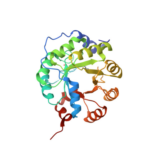
    6TF8, 6TFA

  • PubMed Abstract: 

    Controlling regio- and stereoselectivity of aldol additions is generally challenging. Here we show that an artificial aldolase with high specificity for acetone as the aldol donor can be reengineered via single active site mutations to accept linear and cyclic aliphatic ketones with notable efficiency, regioselectivity, and stereocontrol. Biochemical and crystallographic data show how the mutated residues modulate the binding and activation of specific aldol donors, as well as their subsequent reaction with diverse aldehyde acceptors. Broadening the substrate scope of this evolutionarily naïve catalyst proved much easier than previous attempts to redesign natural aldolases, suggesting that such proteins may be excellent starting points for the development of customized biocatalysts for diverse practical applications.


  • Organizational Affiliation
    • Laboratory of Organic Chemistry, ETH Zurich, 8093 Zurich, Switzerland.

Macromolecule Content 

  • Total Structure Weight: 116.25 kDa 
  • Atom Count: 8,411 
  • Modeled Residue Count: 986 
  • Deposited Residue Count: 1,004 
  • Unique protein chains: 1

Macromolecules

Find similar proteins by:|  3D Structure
Entity ID: 1
MoleculeChains  Sequence LengthOrganismDetailsImage
RA95.5-8F_133F
A, B, C, D
251Saccharolobus solfataricus P2Mutation(s): 0 

Small Molecules

Modified Residues  1 Unique
IDChains TypeFormula2D DiagramParent
N65
Query on N65
A, B, C, D
L-PEPTIDE LINKINGC11 H22 N2 O2LYS

Experimental Data & Validation

Experimental Data

  • Method: X-RAY DIFFRACTION
  • Resolution: 1.90 Å
  • R-Value Free:  0.247 (Depositor), 0.248 (DCC) 
  • R-Value Work:  0.200 (Depositor), 0.200 (DCC) 
  • R-Value Observed: 0.201 (Depositor) 
Space Group: P 21 21 21
Unit Cell:
Length ( Å )Angle ( ˚ )
a = 75.885α = 90
b = 102.316β = 90
c = 121.076γ = 90
Software Package:
Software NamePurpose
PHENIXrefinement
XSCALEdata scaling
PDB_EXTRACTdata extraction
XDSdata reduction
PHASERphasing

Structure Validation

View Full Validation Report


Entry History 

Deposition Data

Revision History  (Full details and data files)

  • Version 1.0: 2020-06-03
    Type: Initial release
  • Version 1.1: 2020-06-17
    Changes: Database references
  • Version 1.2: 2024-01-24
    Changes: Data collection, Database references, Derived calculations, Refinement description
  • Version 1.3: 2024-10-23
    Changes: Structure summary