6TEP | pdb_00006tep

Crystal structure of a galactokinase from Bifidobacterium infantis in complex with ADP


Experimental Data Snapshot

  • Method: X-RAY DIFFRACTION
  • Resolution: 1.45 Å
  • R-Value Free: 
    0.245 (Depositor), 0.248 (DCC) 
  • R-Value Work: 
    0.184 (Depositor), 0.189 (DCC) 
  • R-Value Observed: 
    0.187 (Depositor) 

Starting Model: experimental
View more details

wwPDB Validation 3D Report Full Report

Validation slider image for 6TEP

Ligand Structure Quality Assessment 


This is version 1.3 of the entry. See complete history

Literature

Profiling Substrate Promiscuity of Wild-Type Sugar Kinases for Multi-fluorinated Monosaccharides.

Keenan, T.Parmeggiani, F.Malassis, J.Fontenelle, C.Q.Vendeville, J.B.Offen, W.Both, P.Huang, K.Marchesi, A.Heyam, A.Young, C.Charnock, S.J.Davies, G.J.Linclau, B.Flitsch, S.L.Fascione, M.A.

(2020) Cell Chem Biol 27: 1199

  • DOI: https://doi.org/10.1016/j.chembiol.2020.06.005
  • Primary Citation Related Structures: 
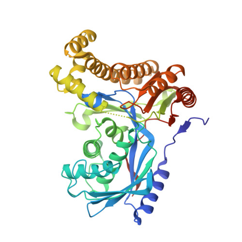
    6TEP, 6TEQ, 6TER

  • PubMed Abstract: 

    Fluorinated sugar-1-phosphates are of emerging importance as intermediates in the chemical and biocatalytic synthesis of modified oligosaccharides, as well as probes for chemical biology. Here we present a systematic study of the activity of a wide range of anomeric sugar kinases (galacto- and N-acetylhexosamine kinases) against a panel of fluorinated monosaccharides, leading to the first examples of polyfluorinated substrates accepted by this class of enzymes. We have discovered four new N-acetylhexosamine kinases with a different substrate scope, thus expanding the number of homologs available in this subclass of kinases. Lastly, we have solved the crystal structure of a galactokinase in complex with 2-deoxy-2-fluorogalactose, giving insight into changes in the active site that may account for the specificity of the enzyme toward certain substrate analogs.


  • Organizational Affiliation
    • Department of Chemistry, University of York, Heslington, York YO10 5DD, UK.

Macromolecule Content 

  • Total Structure Weight: 187.44 kDa 
  • Atom Count: 14,438 
  • Modeled Residue Count: 1,658 
  • Deposited Residue Count: 1,716 
  • Unique protein chains: 1

Macromolecules

Find similar proteins by:|  3D Structure
Entity ID: 1
MoleculeChains  Sequence LengthOrganismDetailsImage
GalactokinaseA,
B [auth D],
C,
D [auth B]
429Bifidobacterium longum subsp. infantis ATCC 15697 = JCM 1222 = DSM 20088Mutation(s): 0 
Gene Names: Blon_2062
EC: 2.7.1.6
UniProt
Find proteins for B7GUI0 (Bifidobacterium longum subsp. infantis (strain ATCC 15697 / DSM 20088 / JCM 1222 / NCTC 11817 / S12))
Explore B7GUI0 
Go to UniProtKB:  B7GUI0
Entity Groups
Sequence Clusters30% Identity50% Identity70% Identity90% Identity95% Identity100% Identity
UniProt GroupB7GUI0
Sequence Annotations
Expand
Reference Sequence

Small Molecules

Ligands 6 Unique
IDChains Name / Formula / InChI Key2D Diagram3D Interactions
ADP
(Subject of Investigation/LOI)

Query on ADP


Download:Ideal Coordinates CCD File
H [auth A],
JA [auth B],
P [auth D],
Y [auth C]
ADENOSINE-5'-DIPHOSPHATE
C10 H15 N5 O10 P2
XTWYTFMLZFPYCI-KQYNXXCUSA-N
PG4

Query on PG4


Download:Ideal Coordinates CCD File
S [auth C]TETRAETHYLENE GLYCOL
C8 H18 O5
UWHCKJMYHZGTIT-UHFFFAOYSA-N
PEG

Query on PEG


Download:Ideal Coordinates CCD File
BA [auth B]
CA [auth B]
DA [auth B]
E [auth A]
EA [auth B]
BA [auth B],
CA [auth B],
DA [auth B],
E [auth A],
EA [auth B],
FA [auth B],
L [auth D],
M [auth D],
N [auth D],
T [auth C],
U [auth C]
DI(HYDROXYETHYL)ETHER
C4 H10 O3
MTHSVFCYNBDYFN-UHFFFAOYSA-N
GOL

Query on GOL


Download:Ideal Coordinates CCD File
F [auth A]
G [auth A]
GA [auth B]
HA [auth B]
IA [auth B]
F [auth A],
G [auth A],
GA [auth B],
HA [auth B],
IA [auth B],
O [auth D],
V [auth C],
W [auth C],
X [auth C]
GLYCEROL
C3 H8 O3
PEDCQBHIVMGVHV-UHFFFAOYSA-N
CL

Query on CL


Download:Ideal Coordinates CCD File
I [auth A],
J [auth A],
KA [auth B],
Q [auth D],
Z [auth C]
CHLORIDE ION
Cl
VEXZGXHMUGYJMC-UHFFFAOYSA-M
MG

Query on MG


Download:Ideal Coordinates CCD File
AA [auth C],
K [auth A],
LA [auth B],
R [auth D]
MAGNESIUM ION
Mg
JLVVSXFLKOJNIY-UHFFFAOYSA-N

Experimental Data & Validation

Experimental Data

  • Method: X-RAY DIFFRACTION
  • Resolution: 1.45 Å
  • R-Value Free:  0.245 (Depositor), 0.248 (DCC) 
  • R-Value Work:  0.184 (Depositor), 0.189 (DCC) 
  • R-Value Observed: 0.187 (Depositor) 
Space Group: P 1 21 1
Unit Cell:
Length ( Å )Angle ( ˚ )
a = 52.272α = 90
b = 164.495β = 95.8
c = 115.873γ = 90
Software Package:
Software NamePurpose
REFMACrefinement
PDB_EXTRACTdata extraction
DIALSdata reduction
Aimlessdata scaling
PHASERphasing

Structure Validation

View Full Validation Report


Ligand Structure Quality Assessment 


Entry History 

& Funding Information

Deposition Data


Funding OrganizationLocationGrant Number
Biotechnology and Biological Sciences Research CouncilUnited KingdomBB/M02847X/1
Biotechnology and Biological Sciences Research CouncilUnited KingdomBB/M028836/1
Biotechnology and Biological Sciences Research CouncilUnited KingdomBB/M028941/1

Revision History  (Full details and data files)

  • Version 1.0: 2020-06-10
    Type: Initial release
  • Version 1.1: 2020-07-22
    Changes: Database references
  • Version 1.2: 2020-09-30
    Changes: Database references, Derived calculations
  • Version 1.3: 2024-01-24
    Changes: Data collection, Database references, Refinement description