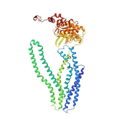
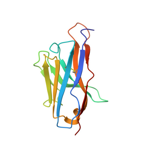
6TEJ

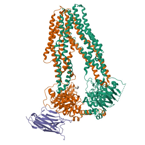
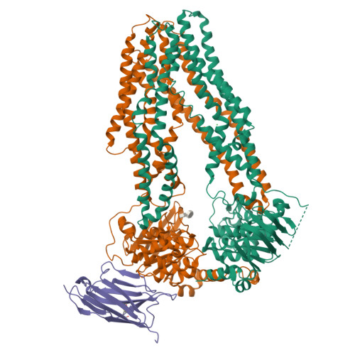
Structure of apo IrtAB devoid SID in complex with sybody Syb_NL5


Experimental Data Snapshot

  • Method: X-RAY DIFFRACTION
  • Resolution: 2.70 Å
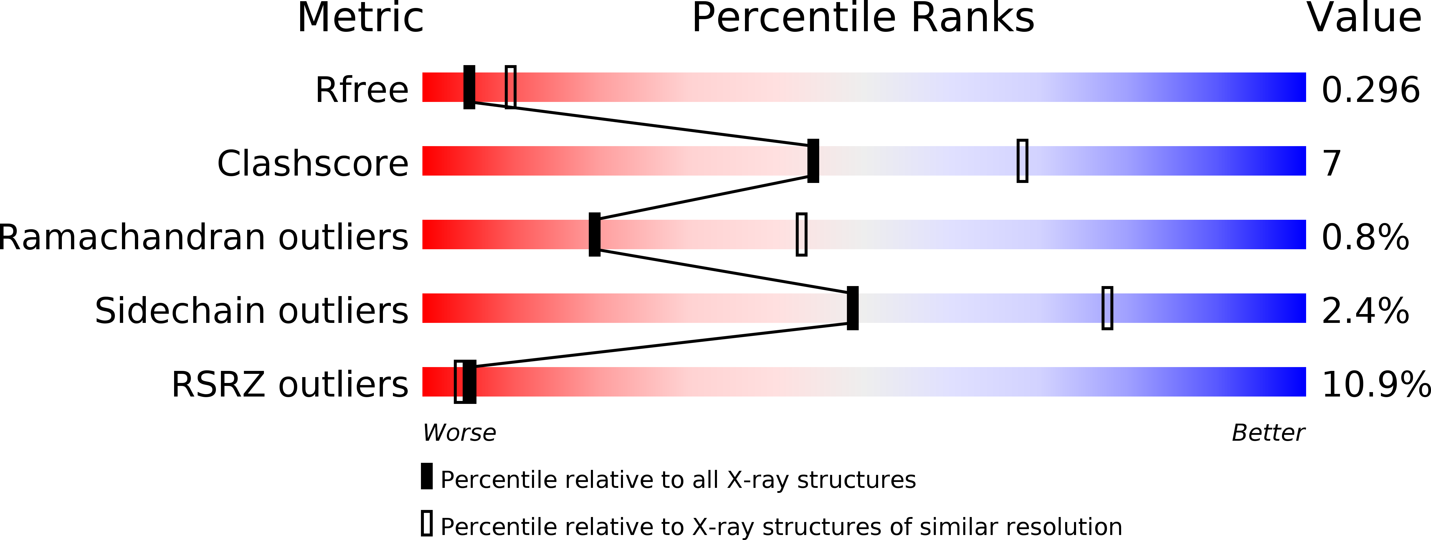
  • R-Value Free: 0.296 
  • R-Value Work: 0.264 
  • R-Value Observed: 0.266 

Starting Model: experimental
View more details

wwPDB Validation   3D Report Full Report


Ligand Structure Quality Assessment 


This is version 1.4 of the entry. See complete history


Literature

The ABC exporter IrtAB imports and reduces mycobacterial siderophores.

Arnold, F.M.Weber, M.S.Gonda, I.Gallenito, M.J.Adenau, S.Egloff, P.Zimmermann, I.Hutter, C.A.J.Hurlimann, L.M.Peters, E.E.Piel, J.Meloni, G.Medalia, O.Seeger, M.A.

(2020) Nature 580: 413-417

  • DOI: https://doi.org/10.1038/s41586-020-2136-9
  • Primary Citation of Related Structures:  
    6TEJ, 6TEK

  • PubMed Abstract: 

    Intracellular replication of the deadly pathogen Mycobacterium tuberculosis relies on the production of small organic molecules called siderophores that scavenge iron from host proteins 1 . M. tuberculosis produces two classes of siderophore, lipid-bound mycobactin and water-soluble carboxymycobactin 2,3 . Functional studies have revealed that iron-loaded carboxymycobactin is imported into the cytoplasm by the ATP binding cassette (ABC) transporter IrtAB 4 , which features an additional cytoplasmic siderophore interaction domain 5 . However, the predicted ABC exporter fold of IrtAB is seemingly contradictory to its import function. Here we show that membrane-reconstituted IrtAB is sufficient to import mycobactins, which are then reduced by the siderophore interaction domain to facilitate iron release. Structure determination by X-ray crystallography and cryo-electron microscopy not only confirms that IrtAB has an ABC exporter fold, but also reveals structural peculiarities at the transmembrane region of IrtAB that result in a partially collapsed inward-facing substrate-binding cavity. The siderophore interaction domain is positioned in close proximity to the inner membrane leaflet, enabling the reduction of membrane-inserted mycobactin. Enzymatic ATPase activity and in vivo growth assays show that IrtAB has a preference for mycobactin over carboxymycobactin as its substrate. Our study provides insights into an unusual ABC exporter that evolved as highly specialized siderophore-import machinery in mycobacteria.


  • Organizational Affiliation

    Institute of Medical Microbiology, University of Zurich, Zurich, Switzerland.


Macromolecules
Find similar proteins by:  (by identity cutoff)  |  3D Structure
Entity ID: 1
MoleculeChains Sequence LengthOrganismDetailsImage
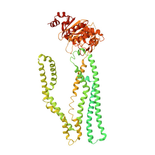
Drug ABC transporter ATP-binding protein908Mycolicibacterium thermoresistibileMutation(s): 0 
Gene Names: RMCT_1990
Membrane Entity: Yes 
UniProt
Find proteins for A0A100XEC2 (Mycolicibacterium thermoresistibile)
Explore A0A100XEC2 
Go to UniProtKB:  A0A100XEC2
Entity Groups  
Sequence Clusters30% Identity50% Identity70% Identity90% Identity95% Identity100% Identity
UniProt GroupA0A100XEC2
Sequence Annotations
Expand
  • Reference Sequence
Find similar proteins by:  (by identity cutoff)  |  3D Structure
Entity ID: 2
MoleculeChains Sequence LengthOrganismDetailsImage
Drug ABC transporter ATP-binding protein586Mycolicibacterium thermoresistibileMutation(s): 0 
Gene Names: RMCT_1989
Membrane Entity: Yes 
UniProt
Find proteins for A0A100XE85 (Mycolicibacterium thermoresistibile)
Explore A0A100XE85 
Go to UniProtKB:  A0A100XE85
Entity Groups  
Sequence Clusters30% Identity50% Identity70% Identity90% Identity95% Identity100% Identity
UniProt GroupA0A100XE85
Sequence Annotations
Expand
  • Reference Sequence
Find similar proteins by:  (by identity cutoff)  |  3D Structure
Entity ID: 3
MoleculeChains Sequence LengthOrganismDetailsImage
Syb_NL5124synthetic constructMutation(s): 0 
Entity Groups  
Sequence Clusters30% Identity50% Identity70% Identity90% Identity95% Identity100% Identity
Sequence Annotations
Expand
  • Reference Sequence
Experimental Data & Validation

Experimental Data

  • Method: X-RAY DIFFRACTION
  • Resolution: 2.70 Å
  • R-Value Free: 0.296 
  • R-Value Work: 0.264 
  • R-Value Observed: 0.266 
  • Space Group: P 1 21 1
Unit Cell:
Length ( Å )Angle ( ˚ )
a = 103.06α = 90
b = 78.58β = 98.2
c = 133.57γ = 90
Software Package:
Software NamePurpose
PHENIXrefinement
Cootmodel building
XSCALEdata reduction
XSCALEdata scaling
PHASERphasing
XDSdata collection

Structure Validation

View Full Validation Report


Ligand Structure Quality Assessment 


Entry History & Funding Information

Deposition Data


Funding OrganizationLocationGrant Number
European Research Council (ERC)Switzerland772190
Swiss National Science FoundationSwitzerlandPP00P3_144823
University of ZurichSwitzerlandK-42225-05-01

Revision History  (Full details and data files)

  • Version 1.0: 2020-04-01
    Type: Initial release
  • Version 1.1: 2020-04-29
    Changes: Database references
  • Version 1.2: 2020-07-01
    Changes: Data collection
  • Version 1.3: 2024-01-24
    Changes: Data collection, Database references, Refinement description
  • Version 1.4: 2024-11-06
    Changes: Structure summary