6TBZ

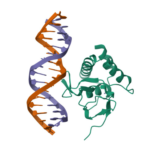
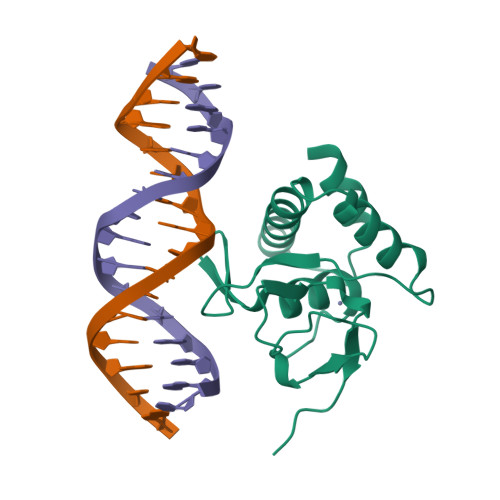
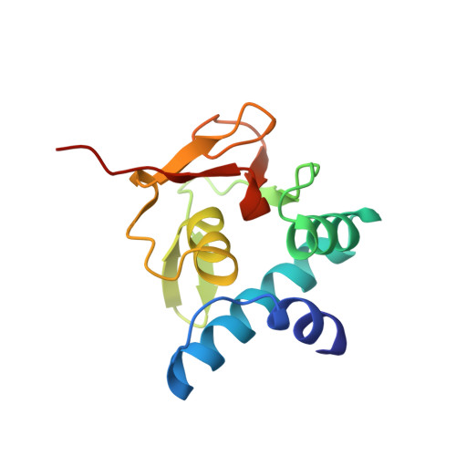
Crystal structure of the MH1 domain of Smad5-Smad3 chimera construct bound to the GGCGC site


Experimental Data Snapshot

  • Method: X-RAY DIFFRACTION
  • Resolution: 1.78 Å
  • R-Value Free: 0.226 
  • R-Value Work: 0.192 
  • R-Value Observed: 0.193 

Starting Model: experimental
View more details

wwPDB Validation   3D Report Full Report


This is version 1.4 of the entry. See complete history


Literature

Unveiling the dimer/monomer propensities of Smad MH1-DNA complexes.

Ruiz, L.Kaczmarska, Z.Gomes, T.Aragon, E.Torner, C.Freier, R.Baginski, B.Martin-Malpartida, P.de Martin Garrido, N.Marquez, J.A.Cordeiro, T.N.Pluta, R.Macias, M.J.

(2021) Comput Struct Biotechnol J 19: 632-646

  • DOI: https://doi.org/10.1016/j.csbj.2020.12.044
  • Primary Citation of Related Structures:  
    6TBZ, 6TCE, 6ZMN

  • PubMed Abstract: 

    Smad transcription factors are the main downstream effectors of the Transforming growth factor β superfamily (TGFβ) signalling network. The DNA complexes determined here by X-ray crystallography for the Bone Morphogenetic Proteins (BMP) activated Smad5 and Smad8 proteins reveal that all MH1 domains bind [GGC(GC)|(CG)] motifs similarly, although TGFβ-activated Smad2/3 and Smad4 MH1 domains bind as monomers whereas Smad1/5/8 form helix-swapped dimers. Dimers and monomers are also present in solution, as revealed by NMR. To decipher the characteristics that defined these dimers, we designed chimeric MH1 domains and characterized them using X-ray crystallography. We found that swapping the loop1 between TGFβ- and BMP- activated MH1 domains switches the dimer/monomer propensities. When we scanned the distribution of Smad-bound motifs in ChIP-Seq peaks (Chromatin immunoprecipitation followed by high-throughput sequencing) in Smad-responsive genes, we observed specific site clustering and spacing depending on whether the peaks correspond to BMP- or TGFβ-responsive genes. We also identified significant correlations between site distribution and monomer or dimer propensities. We propose that the MH1 monomer or dimer propensity of Smads contributes to the distinct motif selection genome-wide and together with the MH2 domain association, help define the composition of R-Smad/Smad4 trimeric complexes.


  • Organizational Affiliation

    Institute for Research in Biomedicine, The Barcelona Institute of Science and Technology, Baldiri Reixac, 10, Barcelona 08028, Spain.


Macromolecules

Find similar proteins by:  (by identity cutoff)  |  3D Structure
Entity ID: 1
MoleculeChains Sequence LengthOrganismDetailsImage
Mothers against decapentaplegic homolog 5131Homo sapiensMutation(s): 0 
Gene Names: SMAD5MADH5
UniProt
Find proteins for Q56I99 (Gallus gallus)
Explore Q56I99 
Go to UniProtKB:  Q56I99
Entity Groups  
Sequence Clusters30% Identity50% Identity70% Identity90% Identity95% Identity100% Identity
UniProt GroupQ56I99
Sequence Annotations
Expand
  • Reference Sequence

Find similar nucleic acids by:  Sequence   |   3D Structure  

Entity ID: 2
MoleculeChains LengthOrganismImage
DNA (5'-D(P*TP*GP*CP*AP*GP*GP*CP*GP*CP*GP*CP*CP*TP*GP*CP*A)-3')
B, C
16Homo sapiens
Sequence Annotations
Expand
  • Reference Sequence
Small Molecules
Ligands 1 Unique
IDChains Name / Formula / InChI Key2D Diagram3D Interactions
ZN (Subject of Investigation/LOI)
Query on ZN

Download Ideal Coordinates CCD File 
D [auth A]ZINC ION
Zn
PTFCDOFLOPIGGS-UHFFFAOYSA-N
Experimental Data & Validation

Experimental Data

  • Method: X-RAY DIFFRACTION
  • Resolution: 1.78 Å
  • R-Value Free: 0.226 
  • R-Value Work: 0.192 
  • R-Value Observed: 0.193 
  • Space Group: P 32
Unit Cell:
Length ( Å )Angle ( ˚ )
a = 53.145α = 90
b = 53.145β = 90
c = 83.154γ = 120
Software Package:
Software NamePurpose
BUSTERrefinement
XDSdata reduction
autoPROCdata scaling
PHASERphasing

Structure Validation

View Full Validation Report


Entry History & Funding Information

Deposition Data


Funding OrganizationLocationGrant Number
Spanish Ministry of Economy and CompetitivenessSpain--
European Commission--

Revision History  (Full details and data files)

  • Version 1.0: 2019-11-20
    Type: Initial release
  • Version 1.1: 2019-12-11
    Changes: Database references, Source and taxonomy, Structure summary
  • Version 1.2: 2020-04-29
    Changes: Structure summary
  • Version 1.3: 2021-02-10
    Changes: Database references, Refinement description
  • Version 1.4: 2024-01-24
    Changes: Advisory, Data collection, Database references, Refinement description