6TAV | pdb_00006tav

Crystal structure of endopeptidase-induced alpha2-macroglobulin


Experimental Data Snapshot

  • Method: X-RAY DIFFRACTION
  • Resolution: 4.20 Å
  • R-Value Free: 
    0.282 (Depositor), 0.329 (DCC) 
  • R-Value Work: 
    0.235 (Depositor) 
  • R-Value Observed: 
    0.236 (Depositor) 

Starting Model: experimental
View more details

wwPDB Validation 3D Report Full Report

Validation slider image for 6TAV

Ligand Structure Quality Assessment 


This is version 1.4 of the entry. See complete history

Literature

Cryo-EM structures show the mechanistic basis of pan-peptidase inhibition by human alpha 2 -macroglobulin.

Luque, D.Goulas, T.Mata, C.P.Mendes, S.R.Gomis-Ruth, F.X.Caston, J.R.

(2022) Proc Natl Acad Sci U S A 119: e2200102119-e2200102119

  • DOI: https://doi.org/10.1073/pnas.2200102119
  • Primary Citation Related Structures: 
    6TAV, 7O7L, 7O7M, 7O7N, 7O7O, 7O7P, 7O7Q, 7O7R, 7O7S

  • PubMed Abstract: 

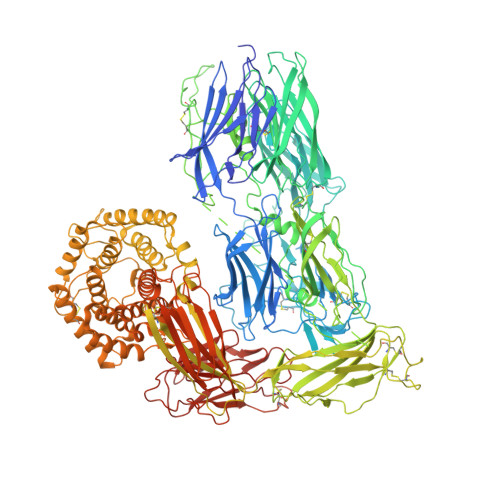
    Human α2-macroglobulin (hα2M) is a multidomain protein with a plethora of essential functions, including transport of signaling molecules and endopeptidase inhibition in innate immunity. Here, we dissected the molecular mechanism of the inhibitory function of the ∼720-kDa hα2M tetramer through eight cryo–electron microscopy (cryo-EM) structures of complexes from human plasma. In the native complex, the hα2M subunits are organized in two flexible modules in expanded conformation, which enclose a highly porous cavity in which the proteolytic activity of circulating plasma proteins is tested. Cleavage of bait regions exposed inside the cavity triggers rearrangement to a compact conformation, which closes openings and entraps the prey proteinase. After the expanded-to-compact transition, which occurs independently in the four subunits, the reactive thioester bond triggers covalent linking of the proteinase, and the receptor-binding domain is exposed on the tetramer surface for receptor-mediated clearance from circulation. These results depict the molecular mechanism of a unique suicidal inhibitory trap.


  • Organizational Affiliation
    • Spanish National Microbiology Centre, Institute of Health Carlos III, 28220 Madrid, Spain.

Macromolecule Content 

  • Total Structure Weight: 666.2 kDa 
  • Atom Count: 41,633 
  • Modeled Residue Count: 5,234 
  • Deposited Residue Count: 5,896 
  • Unique protein chains: 1

Macromolecules

Find similar proteins by:|  3D Structure
Entity ID: 1
MoleculeChains  Sequence LengthOrganismDetailsImage
Alpha-2-macroglobulin
A, B, C, D
1,474Homo sapiensMutation(s): 0 
Gene Names: A2MCPAMD5FWP007
UniProt & NIH Common Fund Data Resources
Find proteins for P01023 (Homo sapiens)
Explore P01023 
Go to UniProtKB:  P01023
PHAROS:  P01023
GTEx:  ENSG00000175899 
Entity Groups
Sequence Clusters30% Identity50% Identity70% Identity90% Identity95% Identity100% Identity
UniProt GroupP01023
Glycosylation
Glycosylation Sites: 7Go to GlyGen: P01023-1
Sequence Annotations
Expand
Reference Sequence

Oligosaccharides

Help  
Entity ID: 2
MoleculeChains Length2D Diagram GlycosylationD Interactions
alpha-D-mannopyranose-(1-6)-beta-D-mannopyranose-(1-4)-2-acetamido-2-deoxy-beta-D-glucopyranose-(1-4)-2-acetamido-2-deoxy-beta-D-glucopyranose
E, G, J, O, V
4N-Glycosylation
Glycosylation Resources
GlyTouCan: G22573RC
GlyCosmos: G22573RC
GlyGen: G22573RC
Entity ID: 3
MoleculeChains Length2D Diagram GlycosylationD Interactions
beta-D-mannopyranose-(1-4)-2-acetamido-2-deoxy-beta-D-glucopyranose-(1-4)-2-acetamido-2-deoxy-beta-D-glucopyranose
F, I, M, R
3N-Glycosylation
Glycosylation Resources
GlyTouCan: G15407YE
GlyCosmos: G15407YE
GlyGen: G15407YE
Entity ID: 4
MoleculeChains Length2D Diagram GlycosylationD Interactions
2-acetamido-2-deoxy-beta-D-glucopyranose-(1-4)-2-acetamido-2-deoxy-beta-D-glucopyranose
H, K, L, N, P
H, K, L, N, P, Q, S, T, U
2N-Glycosylation
Glycosylation Resources
GlyTouCan: G42666HT
GlyCosmos: G42666HT
GlyGen: G42666HT

Small Molecules

Ligands 1 Unique
IDChains Name / Formula / InChI Key2D Diagram3D Interactions
NAG

Query on NAG


Download:Ideal Coordinates CCD File
AA [auth C]
BA [auth C]
CA [auth C]
DA [auth C]
EA [auth D]
AA [auth C],
BA [auth C],
CA [auth C],
DA [auth C],
EA [auth D],
FA [auth D],
GA [auth D],
W [auth A],
X [auth A],
Y [auth A],
Z [auth B]
2-acetamido-2-deoxy-beta-D-glucopyranose
C8 H15 N O6
OVRNDRQMDRJTHS-FMDGEEDCSA-N

Experimental Data & Validation

Experimental Data

  • Method: X-RAY DIFFRACTION
  • Resolution: 4.20 Å
  • R-Value Free:  0.282 (Depositor), 0.329 (DCC) 
  • R-Value Work:  0.235 (Depositor) 
  • R-Value Observed: 0.236 (Depositor) 
Space Group: P 21 21 21
Unit Cell:
Length ( Å )Angle ( ˚ )
a = 130.8α = 90
b = 260.3β = 90
c = 281.8γ = 90
Software Package:
Software NamePurpose
BUSTERrefinement
XSCALEdata scaling
PDB_EXTRACTdata extraction
XDSdata reduction
BUSTERphasing

Structure Validation

View Full Validation Report


Ligand Structure Quality Assessment 


Entry History 

Deposition Data

Revision History  (Full details and data files)

  • Version 1.0: 2021-05-12
    Type: Initial release
  • Version 1.1: 2022-04-13
    Changes: Advisory, Database references
  • Version 1.2: 2024-01-24
    Changes: Data collection, Refinement description
  • Version 1.3: 2024-11-13
    Changes: Structure summary
  • Version 1.4: 2025-09-17
    Changes: Database references