6T8I | pdb_00006t8i

Crystal structure of wild type EndoBT-3987 from Bacteroides thetaiotamicron VPI-5482


Experimental Data Snapshot

  • Method: X-RAY DIFFRACTION
  • Resolution: 1.40 Å
  • R-Value Free: 
    0.181 (Depositor), 0.185 (DCC) 
  • R-Value Work: 
    0.163 (Depositor), 0.166 (DCC) 
  • R-Value Observed: 
    0.164 (Depositor) 

Starting Model: experimental
View more details

wwPDB Validation 3D Report Full Report

Validation slider image for 6T8I

This is version 1.1 of the entry. See complete history

Literature

Structural basis of mammalian high-mannose N-glycan processing by human gut Bacteroides.

Trastoy, B.Du, J.J.Klontz, E.H.Li, C.Cifuente, J.O.Wang, L.X.Sundberg, E.J.Guerin, M.E.

(2020) Nat Commun 11: 899-899

  • DOI: https://doi.org/10.1038/s41467-020-14754-7
  • Primary Citation Related Structures: 
    6T8I, 6T8K, 6T8L, 6TCV, 6TCW

  • PubMed Abstract: 

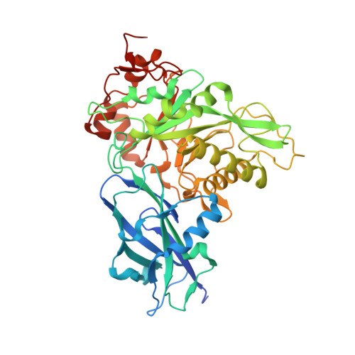
    The human gut microbiota plays a central role not only in regulating the metabolism of nutrients but also promoting immune homeostasis, immune responses and protection against pathogen colonization. The genome of the Gram-negative symbiont Bacteroides thetaiotaomicron, a dominant member of the human intestinal microbiota, encodes polysaccharide utilization loci PULs, the apparatus required to orchestrate the degradation of a specific glycan. EndoBT-3987 is a key endo-β-N-acetylglucosaminidase (ENGase) that initiates the degradation/processing of mammalian high-mannose-type (HM-type) N-glycans in the intestine. Here, we provide structural snapshots of EndoBT-3987, including the unliganded form, the EndoBT-3987-Man 9 GlcNAc 2 Asn substrate complex, and two EndoBT-3987-Man 9 GlcNAc and EndoBT-3987-Man 5 GlcNAc product complexes. In combination with alanine scanning mutagenesis and activity measurements we unveil the molecular mechanism of HM-type recognition and specificity for EndoBT-3987 and an important group of the GH18 ENGases, including EndoH, an enzyme extensively used in biotechnology, and for which the mechanism of substrate recognition was largely unknown.


  • Organizational Affiliation
    • Structural Biology Unit, Center for Cooperative Research in Biosciences (CIC bioGUNE), Basque Research and Technology Alliance (BRTA), Bizkaia Technology Park, Building 801A, 48160, Derio, Spain. btrastoy@cicbiogune.es.

Macromolecule Content 

  • Total Structure Weight: 49.38 kDa 
  • Atom Count: 3,692 
  • Modeled Residue Count: 434 
  • Deposited Residue Count: 451 
  • Unique protein chains: 1

Macromolecules

Find similar proteins by:|  3D Structure
Entity ID: 1
MoleculeChains  Sequence LengthOrganismDetailsImage
Endo-beta-N-acetylglucosaminidase F1451Bacteroides thetaiotaomicron VPI-5482Mutation(s): 0 
Gene Names: BT_3987
UniProt
Find proteins for Q8A0N4 (Bacteroides thetaiotaomicron (strain ATCC 29148 / DSM 2079 / JCM 5827 / CCUG 10774 / NCTC 10582 / VPI-5482 / E50))
Explore Q8A0N4 
Go to UniProtKB:  Q8A0N4
Entity Groups
Sequence Clusters30% Identity50% Identity70% Identity90% Identity95% Identity100% Identity
UniProt GroupQ8A0N4
Sequence Annotations
Expand
Reference Sequence

Experimental Data & Validation

Experimental Data

  • Method: X-RAY DIFFRACTION
  • Resolution: 1.40 Å
  • R-Value Free:  0.181 (Depositor), 0.185 (DCC) 
  • R-Value Work:  0.163 (Depositor), 0.166 (DCC) 
  • R-Value Observed: 0.164 (Depositor) 
Space Group: P 31 2 1
Unit Cell:
Length ( Å )Angle ( ˚ )
a = 74.578α = 90
b = 74.578β = 90
c = 133.54γ = 120
Software Package:
Software NamePurpose
PHENIXrefinement
PDB_EXTRACTdata extraction
XDSdata reduction
XDSdata scaling
PHENIXphasing

Structure Validation

View Full Validation Report


Entry History 

Deposition Data

Revision History  (Full details and data files)

  • Version 1.0: 2020-02-26
    Type: Initial release
  • Version 1.1: 2024-01-24
    Changes: Data collection, Database references, Refinement description