


Crystal structure of 2-methylisocitrate lyase (PrpB) from Pseudomonas aeruginosa in complex with Mg(II)-pyruvate.
Wijaya, A.J., Brear, P., Dolan, S.K., Welch, M.To be published.
Experimental Data Snapshot
Starting Model: experimental
View more details
Entity ID: 1 | |||||
|---|---|---|---|---|---|
| Molecule | Chains | Sequence Length | Organism | Details | Image |
| 2-methylisocitrate lyase | A [auth C], B [auth A], C [auth D], D [auth B] | 298 | Pseudomonas aeruginosa PAO1 | Mutation(s): 0 Gene Names: prpB, PA0796 EC: 4.1.3.30 | 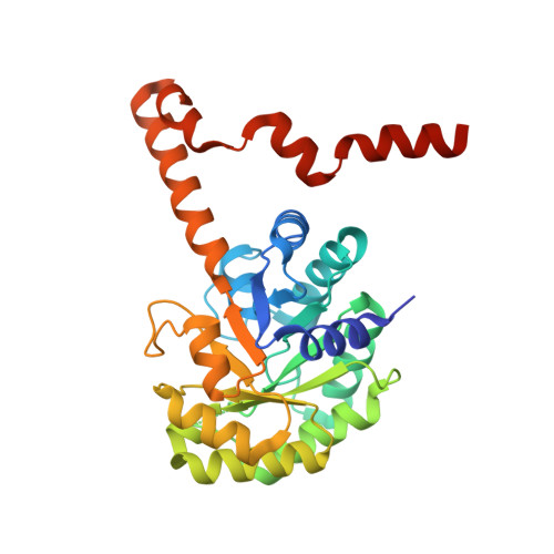 |
UniProt | |||||
Entity Groups | |||||
| Sequence Clusters | 30% Identity50% Identity70% Identity90% Identity95% Identity100% Identity | ||||
| UniProt Group | Q9I5E2 | ||||
Sequence AnnotationsExpand | |||||
Reference Sequence | |||||
| Ligands 2 Unique | |||||
|---|---|---|---|---|---|
| ID | Chains | Name / Formula / InChI Key | 2D Diagram | 3D Interactions | |
| PYR (Subject of Investigation/LOI) Download:Ideal Coordinates CCD File | F [auth C], H [auth A], J [auth D], L [auth B] | PYRUVIC ACID C3 H4 O3 LCTONWCANYUPML-UHFFFAOYSA-N |  | ||
| MG (Subject of Investigation/LOI) Download:Ideal Coordinates CCD File | E [auth C], G [auth A], I [auth D], K [auth B] | MAGNESIUM ION Mg JLVVSXFLKOJNIY-UHFFFAOYSA-N |  | ||
| Length ( Å ) | Angle ( ˚ ) |
|---|---|
| a = 153.49 | α = 90 |
| b = 59.4 | β = 120.3 |
| c = 147.65 | γ = 90 |
| Software Name | Purpose |
|---|---|
| PHENIX | refinement |
| Aimless | data scaling |
| PDB_EXTRACT | data extraction |
| xia2 | data reduction |
| PHASER | phasing |
| Funding Organization | Location | Grant Number |
|---|---|---|
| Biotechnology and Biological Sciences Research Council | United Kingdom | BB/M019411/1 |
| Biotechnology and Biological Sciences Research Council | United Kingdom | BB/R005435/1 |


