6T3Q | pdb_00006t3q

Thrombin in Complex with a D-Phe-Pro-2-aminopyridine derivative


Experimental Data Snapshot

  • Method: X-RAY DIFFRACTION
  • Resolution: 1.33 Å
  • R-Value Free: 
    0.146 (Depositor), 0.146 (DCC) 
  • R-Value Work: 
    0.125 (Depositor), 0.126 (DCC) 
  • R-Value Observed: 
    0.126 (Depositor) 

Starting Model: experimental
View more details

wwPDB Validation 3D Report Full Report

Validation slider image for 6T3Q

Ligand Structure Quality Assessment 


This is version 1.3 of the entry. See complete history

Literature

Protein-Induced Change in Ligand Protonation during Trypsin and Thrombin Binding: Hint on Differences in Selectivity Determinants of Both Proteins?

Ngo, K.Collins-Kautz, C.Gerstenecker, S.Wagner, B.Heine, A.Klebe, G.

(2020) J Med Chem 63: 3274-3289

  • DOI: https://doi.org/10.1021/acs.jmedchem.9b02061
  • Primary Citation Related Structures: 
    6HSX, 6SY3, 6T0M, 6T0P, 6T3Q, 6T4A, 6T5W, 6TDT

  • PubMed Abstract: 

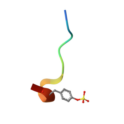
    Trypsin and thrombin, structurally similar serine proteases, recognize different substrates; thrombin cleaves after Arg, whereas trypsin cleaves after Lys/Arg. Both recognize basic substrate headgroups via Asp189 at the bottom of the S1 pocket. By crystallography and isothermal titration calorimetry (ITC), we studied a series of d-Phe/d-DiPhe-Pro-(amino)pyridines. Identical ligand pairs show the same binding poses. Surprisingly, one ligand binds to trypsin in protonated state and to thrombin in unprotonated state at P1 along with differences in the residual solvation pattern. While trypsin binding is mediated by an ordered water molecule, in thrombin, water is scattered over three hydration sites. Although having highly similar S1 pockets, our results suggest different electrostatic properties of Asp189 possibly contributing to the selectivity determinant. Thrombin binds a specific Na + ion next to Asp189, which is absent in trypsin. The electrostatic properties across the S1 pocket are further attenuated by charged Glu192 at the rim of S1 in thrombin, which is replaced by uncharged Gln192 in trypsin.


  • Organizational Affiliation
    • Institute of Pharmaceutical Chemistry, Philipps-University Marburg, Marbacher Weg 6, 35032 Marburg, Germany.

Macromolecule Content 

  • Total Structure Weight: 36.19 kDa 
  • Atom Count: 2,714 
  • Modeled Residue Count: 290 
  • Deposited Residue Count: 306 
  • Unique protein chains: 3

Macromolecules

Find similar proteins by:|  3D Structure
Entity ID: 1
MoleculeChains  Sequence LengthOrganismDetailsImage
ProthrombinA [auth L]36Homo sapiensMutation(s): 0 
EC: 3.4.21.5
UniProt & NIH Common Fund Data Resources
Find proteins for P00734 (Homo sapiens)
Explore P00734 
Go to UniProtKB:  P00734
PHAROS:  P00734
GTEx:  ENSG00000180210 
Entity Groups
Sequence Clusters30% Identity50% Identity70% Identity90% Identity95% Identity100% Identity
UniProt GroupP00734
Sequence Annotations
Expand
Reference Sequence
Find similar proteins by:|  3D Structure
Entity ID: 2
MoleculeChains  Sequence LengthOrganismDetailsImage
ProthrombinB [auth H]259Homo sapiensMutation(s): 0 
EC: 3.4.21.5
UniProt & NIH Common Fund Data Resources
Find proteins for P00734 (Homo sapiens)
Explore P00734 
Go to UniProtKB:  P00734
PHAROS:  P00734
GTEx:  ENSG00000180210 
Entity Groups
Sequence Clusters30% Identity50% Identity70% Identity90% Identity95% Identity100% Identity
UniProt GroupP00734
Glycosylation
Glycosylation Sites: 1Go to GlyGen: P00734-1
Sequence Annotations
Expand
Reference Sequence
Find similar proteins by:  Sequence   |   3D Structure  
Entity ID: 3
MoleculeChains  Sequence LengthOrganismDetailsImage
Hirudin variant-2C [auth I]11Hirudo medicinalisMutation(s): 0 
UniProt
Find proteins for P09945 (Hirudo medicinalis)
Explore P09945 
Go to UniProtKB:  P09945
Entity Groups
Sequence Clusters30% Identity50% Identity70% Identity90% Identity95% Identity100% Identity
UniProt GroupP09945
Sequence Annotations
Expand
Reference Sequence

Small Molecules

Ligands 5 Unique
IDChains Name / Formula / InChI Key2D Diagram3D Interactions
M6Q
(Subject of Investigation/LOI)

Query on M6Q


Download:Ideal Coordinates CCD File
I [auth H](2~{S})-1-[(2~{R})-2-azanyl-3-phenyl-propanoyl]-~{N}-[(2-azanylpyridin-4-yl)methyl]pyrrolidine-2-carboxamide
C20 H25 N5 O2
LOSWCWUZWRHVLI-SJORKVTESA-N
NAG

Query on NAG


Download:Ideal Coordinates CCD File
D [auth H]2-acetamido-2-deoxy-beta-D-glucopyranose
C8 H15 N O6
OVRNDRQMDRJTHS-FMDGEEDCSA-N
PO4

Query on PO4


Download:Ideal Coordinates CCD File
E [auth H]PHOSPHATE ION
O4 P
NBIIXXVUZAFLBC-UHFFFAOYSA-K
GOL

Query on GOL


Download:Ideal Coordinates CCD File
F [auth H]GLYCEROL
C3 H8 O3
PEDCQBHIVMGVHV-UHFFFAOYSA-N
NA

Query on NA


Download:Ideal Coordinates CCD File
G [auth H],
H
SODIUM ION
Na
FKNQFGJONOIPTF-UHFFFAOYSA-N
Modified Residues  1 Unique
IDChains TypeFormula2D DiagramParent
TYS
Query on TYS
C [auth I]L-PEPTIDE LINKINGC9 H11 N O6 STYR
Binding Affinity Annotations 
IDSourceBinding Affinity
M6Q BindingDB:  6T3Q Ki: 379 (nM) from 1 assay(s)
Kd: 916 (nM) from 1 assay(s)

Experimental Data & Validation

Experimental Data

  • Method: X-RAY DIFFRACTION
  • Resolution: 1.33 Å
  • R-Value Free:  0.146 (Depositor), 0.146 (DCC) 
  • R-Value Work:  0.125 (Depositor), 0.126 (DCC) 
  • R-Value Observed: 0.126 (Depositor) 
Space Group: C 1 2 1
Unit Cell:
Length ( Å )Angle ( ˚ )
a = 70.009α = 90
b = 71.29β = 100.585
c = 72.572γ = 90
Software Package:
Software NamePurpose
PHENIXrefinement
HKL-2000data reduction
HKL-2000data scaling
PHASERphasing

Structure Validation

View Full Validation Report


Ligand Structure Quality Assessment 


Entry History 

Deposition Data

Revision History  (Full details and data files)

  • Version 1.0: 2020-05-13
    Type: Initial release
  • Version 1.1: 2020-07-29
    Type: Remediation
    Reason: Carbohydrate remediation
    Changes: Data collection, Derived calculations, Structure summary
  • Version 1.2: 2024-01-24
    Changes: Data collection, Database references, Refinement description, Structure summary
  • Version 1.3: 2024-11-06
    Changes: Structure summary