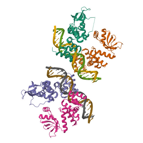
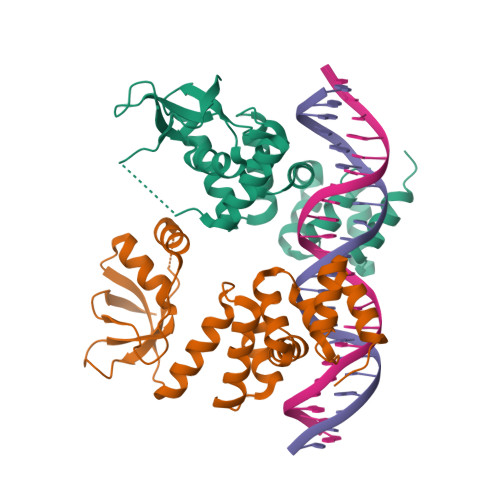
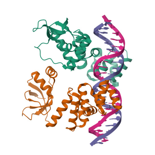
6T1F

Crystal structure of the C-terminally truncated chromosome-partitioning protein ParB from Caulobacter crescentus complexed to the centromeric parS site


Experimental Data Snapshot

  • Method: X-RAY DIFFRACTION
  • Resolution: 2.90 Å
  • R-Value Free: 0.263 
  • R-Value Work: 0.239 
  • R-Value Observed: 0.241 

Starting Model: experimental
View more details

wwPDB Validation   3D Report Full Report


This is version 1.2 of the entry. See complete history


Literature

A CTP-dependent gating mechanism enables ParB spreading on DNA.

Jalal, A.S.Tran, N.T.Stevenson, C.E.Chimthanawala, A.Badrinarayanan, A.Lawson, D.M.Le, T.B.

(2021) Elife 10

  • DOI: https://doi.org/10.7554/eLife.69676
  • Primary Citation of Related Structures:  
    6T1F, 7BM8

  • PubMed Abstract: 

    Proper chromosome segregation is essential in all living organisms. The ParA-ParB- parS system is widely employed for chromosome segregation in bacteria. Previously, we showed that Caulobacter crescentus ParB requires cytidine triphosphate to escape the nucleation site parS and spread by sliding to the neighboring DNA (Jalal et al., 2020). Here, we provide the structural basis for this transition from nucleation to spreading by solving co-crystal structures of a C-terminal domain truncated C. crescentus ParB with parS and with a CTP analog. Nucleating ParB is an open clamp, in which parS is captured at the DNA-binding domain (the DNA-gate). Upon binding CTP, the N-terminal domain (NTD) self-dimerizes to close the NTD-gate of the clamp. The DNA-gate also closes, thus driving parS into a compartment between the DNA-gate and the C-terminal domain. CTP hydrolysis and/or the release of hydrolytic products are likely associated with reopening of the gates to release DNA and recycle ParB. Overall, we suggest a CTP-operated gating mechanism that regulates ParB nucleation, spreading, and recycling.


  • Organizational Affiliation

    Department of Molecular Microbiology, John Innes Centre, Norwich, United Kingdom.


Macromolecules

Find similar proteins by:  (by identity cutoff)  |  3D Structure
Entity ID: 1
MoleculeChains Sequence LengthOrganismDetailsImage
Chromosome-partitioning protein ParB
A, B, C, D
257Caulobacter vibrioides NA1000Mutation(s): 0 
Gene Names: parBCCNA_03868
UniProt
Find proteins for B8GW30 (Caulobacter vibrioides (strain NA1000 / CB15N))
Explore B8GW30 
Go to UniProtKB:  B8GW30
Entity Groups  
Sequence Clusters30% Identity50% Identity70% Identity90% Identity95% Identity100% Identity
UniProt GroupB8GW30
Sequence Annotations
Expand
  • Reference Sequence

Find similar nucleic acids by:  Sequence   |   3D Structure  

Entity ID: 2
MoleculeChains LengthOrganismImage
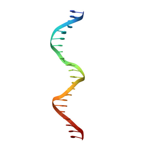
DNA (5'-D(*GP*GP*AP*TP*GP*TP*TP*TP*CP*AP*CP*GP*TP*GP*AP*AP*AP*CP*AP*TP*CP*C)-3')
E, F, G, H
22Caulobacter vibrioides
Sequence Annotations
Expand
  • Reference Sequence
Experimental Data & Validation

Experimental Data

  • Method: X-RAY DIFFRACTION
  • Resolution: 2.90 Å
  • R-Value Free: 0.263 
  • R-Value Work: 0.239 
  • R-Value Observed: 0.241 
  • Space Group: P 1 21 1
Unit Cell:
Length ( Å )Angle ( ˚ )
a = 54.25α = 90
b = 172.928β = 90.54
c = 72.852γ = 90
Software Package:
Software NamePurpose
XDSdata reduction
Aimlessdata scaling
REFMACrefinement
PDB_EXTRACTdata extraction
PHASERphasing

Structure Validation

View Full Validation Report


Entry History & Funding Information

Deposition Data


Funding OrganizationLocationGrant Number
Royal SocietyUnited KingdomUF140053
Royal SocietyUnited KingdomRG150448
Biotechnology and Biological Sciences Research CouncilUnited KingdomBB/P018165/1
Biotechnology and Biological Sciences Research CouncilUnited KingdomBBS/E/J/000C0683
Biotechnology and Biological Sciences Research CouncilUnited KingdomBB/P012523/1

Revision History  (Full details and data files)

  • Version 1.0: 2020-10-14
    Type: Initial release
  • Version 1.1: 2021-10-13
    Changes: Data collection, Database references
  • Version 1.2: 2024-01-24
    Changes: Data collection, Refinement description