6T18

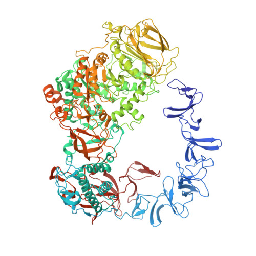
ASR Alternansucrase in complex with oligoalternan


Experimental Data Snapshot

  • Method: X-RAY DIFFRACTION
  • Resolution: 3.15 Å
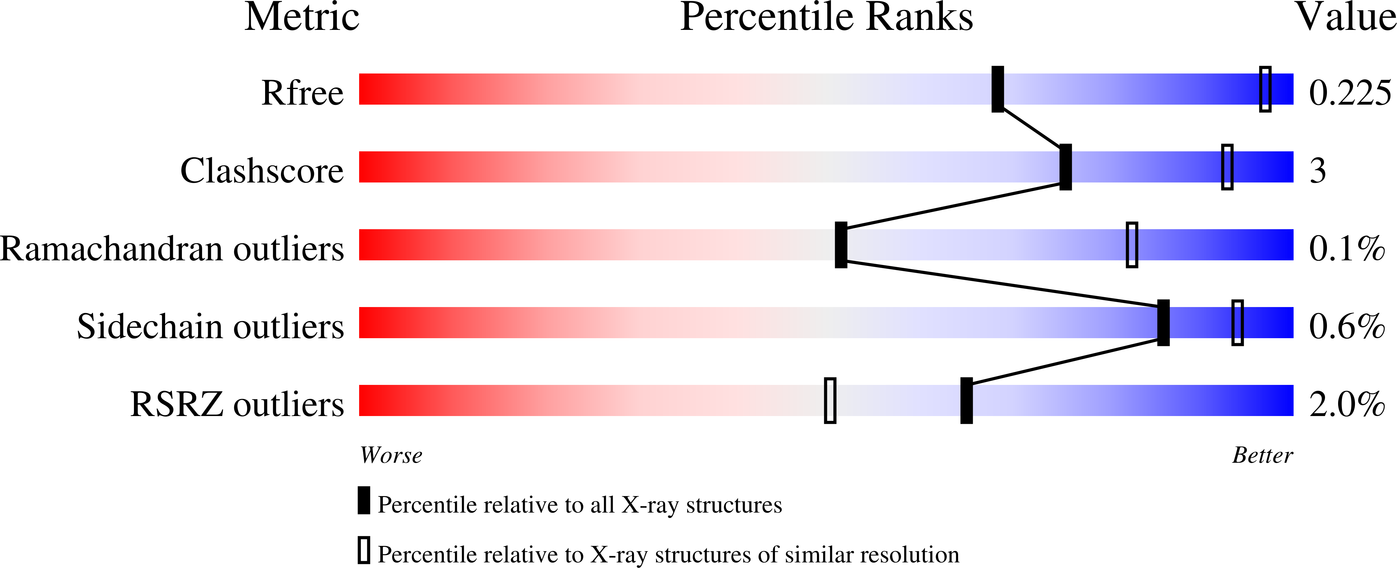
  • R-Value Free: 0.226 
  • R-Value Work: 0.200 
  • R-Value Observed: 0.201 

Starting Model: experimental
View more details

wwPDB Validation   3D Report Full Report


This is version 2.1 of the entry. See complete history


Literature

A specific oligosaccharide-binding site in the alternansucrase catalytic domain mediates alternan elongation.

Molina, M.Moulis, C.Monties, N.Guieysse, D.Morel, S.Cioci, G.Remaud-Simeon, M.

(2020) J Biol Chem 295: 9474-9489

  • DOI: https://doi.org/10.1074/jbc.RA120.013028
  • Primary Citation of Related Structures:  
    6SYQ, 6SZI, 6T16, 6T18, 6T1P

  • PubMed Abstract: 

    Microbial α-glucans produced by GH70 (glycoside hydrolase family 70) glucansucrases are gaining importance because of the mild conditions for their synthesis from sucrose, their biodegradability, and their current and anticipated applications that largely depend on their molar mass. Focusing on the alternansucrase (ASR) from Leuconostoc citreum NRRL B-1355, a well-known glucansucrase catalyzing the synthesis of both high- and low-molar-mass alternans, we searched for structural traits in ASR that could be involved in the control of alternan elongation. The resolution of five crystal structures of a truncated ASR version (ASRΔ2) in complex with different gluco-oligosaccharides pinpointed key residues in binding sites located in the A and V domains of ASR. Biochemical characterization of three single mutants and three double mutants targeting the sugar-binding pockets identified in domain V revealed an involvement of this domain in alternan binding and elongation. More strikingly, we found an oligosaccharide-binding site at the surface of domain A, distant from the catalytic site and not previously identified in other glucansucrases. We named this site surface-binding site (SBS) A1. Among the residues lining the SBS-A1 site, two (Gln 700 and Tyr 717 ) promoted alternan elongation. Their substitution to alanine decreased high-molar-mass alternan yield by a third, without significantly impacting enzyme stability or specificity. We propose that the SBS-A1 site is unique to alternansucrase and appears to be designed to bind alternating structures, acting as a mediator between the catalytic site and the sugar-binding pockets of domain V and contributing to a processive elongation of alternan chains.


  • Organizational Affiliation

    Toulouse Biotechnology Institute (TBI), Université de Toulouse, CNRS, INRAE, INSA, Toulouse, France.


Macromolecules
Find similar proteins by:  (by identity cutoff)  |  3D Structure
Entity ID: 1
MoleculeChains Sequence LengthOrganismDetailsImage
Alternansucrase
A, B
1,278Leuconostoc mesenteroidesMutation(s): 0 
Gene Names: asr
EC: 2.4.1.140 (PDB Primary Data), 2.4.1.5 (UniProt)
UniProt
Find proteins for Q9RE05 (Leuconostoc mesenteroides)
Explore Q9RE05 
Go to UniProtKB:  Q9RE05
Entity Groups  
Sequence Clusters30% Identity50% Identity70% Identity90% Identity95% Identity100% Identity
UniProt GroupQ9RE05
Sequence Annotations
Expand
  • Reference Sequence
Oligosaccharides

Help

Entity ID: 2
MoleculeChains Length2D Diagram Glycosylation3D Interactions
alpha-D-glucopyranose-(1-6)-alpha-D-glucopyranose-(1-3)-alpha-D-glucopyranose-(1-6)-alpha-D-glucopyranose-(1-3)-alpha-D-glucopyranose-(1-6)-alpha-D-glucopyranose
C
6N/A
Glycosylation Resources
GlyTouCan:  G64325VG
GlyCosmos:  G64325VG
GlyGen:  G64325VG
Entity ID: 3
MoleculeChains Length2D Diagram Glycosylation3D Interactions
alpha-D-glucopyranose-(1-3)-alpha-D-glucopyranose-(1-6)-alpha-D-glucopyranose
D
3N/A
Glycosylation Resources
GlyTouCan:  G92776DG
GlyCosmos:  G92776DG
GlyGen:  G92776DG
Entity ID: 4
MoleculeChains Length2D Diagram Glycosylation3D Interactions
alpha-D-glucopyranose-(1-3)-alpha-D-glucopyranose-(1-6)-alpha-D-glucopyranose-(1-3)-alpha-D-glucopyranose
E
4N/A
Glycosylation Resources
GlyTouCan:  G52995QB
GlyCosmos:  G52995QB
GlyGen:  G52995QB
Experimental Data & Validation

Experimental Data

  • Method: X-RAY DIFFRACTION
  • Resolution: 3.15 Å
  • R-Value Free: 0.226 
  • R-Value Work: 0.200 
  • R-Value Observed: 0.201 
  • Space Group: P 21 21 21
Unit Cell:
Length ( Å )Angle ( ˚ )
a = 100.876α = 90
b = 135.654β = 90
c = 239.424γ = 90
Software Package:
Software NamePurpose
REFMACrefinement
PDB_EXTRACTdata extraction
XDSdata reduction
SCALAdata scaling
PHASERphasing

Structure Validation

View Full Validation Report


Entry History 

Deposition Data

Revision History  (Full details and data files)

  • Version 1.0: 2020-06-17
    Type: Initial release
  • Version 1.1: 2020-07-22
    Changes: Database references
  • Version 2.0: 2020-07-29
    Type: Remediation
    Reason: Carbohydrate remediation
    Changes: Advisory, Atomic model, Data collection, Derived calculations, Structure summary
  • Version 2.1: 2024-01-24
    Changes: Data collection, Database references, Refinement description, Structure summary