


The structure of penicillin-binding protein 3 from Yersinia pestis
Pankov, G.To be published.
Experimental Data Snapshot
Starting Model: experimental
View more details
Entity ID: 1 | |||||
|---|---|---|---|---|---|
| Molecule | Chains | Sequence Length | Organism | Details | Image |
| Peptidoglycan D,D-transpeptidase FtsI | 533 | Yersinia pestis | Mutation(s): 0 Gene Names: ftsI, YPO0549 EC: 3.4.16.4 | 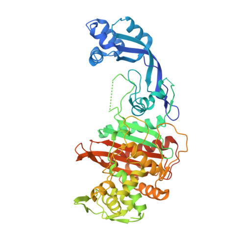 | |
UniProt | |||||
Entity Groups | |||||
| Sequence Clusters | 30% Identity50% Identity70% Identity90% Identity95% Identity100% Identity | ||||
| UniProt Group | A0A3N4B5A3 | ||||
Sequence AnnotationsExpand | |||||
Reference Sequence | |||||
| Ligands 2 Unique | |||||
|---|---|---|---|---|---|
| ID | Chains | Name / Formula / InChI Key | 2D Diagram | 3D Interactions | |
| CB9 Download:Ideal Coordinates CCD File | D [auth A] | (2R,4S)-2-[(1R)-1-{[(2S)-2-carboxy-2-phenylacetyl]amino}-2-oxoethyl]-5,5-dimethyl-1,3-thiazolidine-4-carboxylic acid C17 H20 N2 O6 S YABPSSAFCROUQF-OWTLIXCDSA-N |  | ||
| ACT Download:Ideal Coordinates CCD File | B [auth A], C [auth A] | ACETATE ION C2 H3 O2 QTBSBXVTEAMEQO-UHFFFAOYSA-M |  | ||
| Length ( Å ) | Angle ( ˚ ) |
|---|---|
| a = 40.86 | α = 90 |
| b = 104.95 | β = 90 |
| c = 110.42 | γ = 90 |
| Software Name | Purpose |
|---|---|
| Aimless | data scaling |
| REFMAC | refinement |
| PDB_EXTRACT | data extraction |
| MOSFLM | data reduction |
| PHASER | phasing |


