6SX4 | pdb_00006sx4

Structure of C. glutamicum mycoloyltransferase A


Experimental Data Snapshot

  • Method: X-RAY DIFFRACTION
  • Resolution: 2.80 Å
  • R-Value Free: 
    0.262 (Depositor), 0.266 (DCC) 
  • R-Value Work: 
    0.242 (Depositor), 0.244 (DCC) 

wwPDB Validation 3D Report Full Report

Validation slider image for 6SX4

This is version 1.2 of the entry. See complete history

Literature

The C-terminal domain of Corynebacterium glutamicum mycoloyltransferase A is composed of five repeated motifs involved in cell wall binding and stability.

Dietrich, C.Li de la Sierra-Gallay, I.Masi, M.Girard, E.Dautin, N.Constantinesco-Becker, F.Tropis, M.Daffe, M.van Tilbeurgh, H.Bayan, N.

(2020) Mol Microbiol 114: 1-16

  • DOI: https://doi.org/10.1111/mmi.14492
  • Primary Citation Related Structures: 
    6SWZ, 6SX4

  • PubMed Abstract: 

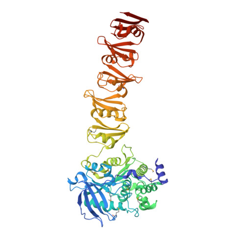
    The genomes of Corynebacteriales contain several genes encoding mycoloyltransferases (Myt) that are specific cell envelope enzymes essential for the biogenesis of the outer membrane. MytA is a major mycoloyltransferase of Corynebacterium glutamicum, displaying an N-terminal domain with esterase activity and a C-terminal extension containing a conserved repeated Leu-Gly-Phe-Pro (LGFP) sequence motif of unknown function. This motif is highly conserved in Corynebacteriales and found associated with cell wall hydrolases and with proteins of unknown function. In this study, we determined the crystal structure of MytA and found that its C-terminal domain is composed of five LGFP motifs and forms a long stalk perpendicular to the N-terminal catalytic α/β-hydrolase domain. The LGFP motifs are composed of a 4-stranded β-fold and occupy alternating orientations along the axis of the stalk. Multiple acetate binding pockets were identified in the stalk, which could correspond to putative ligand-binding sites. By using various MytA mutants and complementary in vitro and in vivo approaches, we provide evidence that the C-terminal LGFP domain interacts with the cell wall peptidoglycan-arabinogalactan polymer. We also show that the C-terminal LGFP domain is not required for the activity of MytA but rather contributes to the overall integrity of the cell envelope.


  • Organizational Affiliation
    • Institute for Integrative Biology of the Cell (I2BC), Université Paris-Saclay, CEA, CNRS, Gif-sur-Yvette, France.

Macromolecule Content 

  • Total Structure Weight: 271.51 kDa 
  • Atom Count: 18,365 
  • Modeled Residue Count: 2,357 
  • Deposited Residue Count: 2,480 
  • Unique protein chains: 1

Macromolecules

Find similar proteins by:|  3D Structure
Entity ID: 1
MoleculeChains  Sequence LengthOrganismDetailsImage
Protein PS1A [auth AAA],
B [auth BBB],
C [auth CCC],
D [auth DDD]
620Corynebacterium glutamicumMutation(s): 0 
Gene Names: csp1cop1Cgl2875cg3182
UniProt
Find proteins for P0C1D6 (Corynebacterium glutamicum (strain ATCC 13032 / DSM 20300 / JCM 1318 / BCRC 11384 / CCUG 27702 / LMG 3730 / NBRC 12168 / NCIMB 10025 / NRRL B-2784 / 534))
Explore P0C1D6 
Go to UniProtKB:  P0C1D6
Entity Groups
Sequence Clusters30% Identity50% Identity70% Identity90% Identity95% Identity100% Identity
UniProt GroupP0C1D6
Sequence Annotations
Expand
Reference Sequence

Small Molecules

Ligands 1 Unique
IDChains Name / Formula / InChI Key2D Diagram3D Interactions
ACT

Query on ACT


Download:Ideal Coordinates CCD File
AA [auth DDD]
BA [auth DDD]
CA [auth DDD]
E [auth AAA]
F [auth AAA]
AA [auth DDD],
BA [auth DDD],
CA [auth DDD],
E [auth AAA],
F [auth AAA],
G [auth AAA],
H [auth AAA],
I [auth AAA],
J [auth AAA],
K [auth AAA],
L [auth BBB],
M [auth BBB],
N [auth BBB],
O [auth BBB],
P [auth BBB],
Q [auth CCC],
R [auth CCC],
S [auth CCC],
T [auth CCC],
U [auth CCC],
V [auth CCC],
W [auth DDD],
X [auth DDD],
Y [auth DDD],
Z [auth DDD]
ACETATE ION
C2 H3 O2
QTBSBXVTEAMEQO-UHFFFAOYSA-M
Modified Residues  1 Unique
IDChains TypeFormula2D DiagramParent
MSE
Query on MSE
A [auth AAA],
B [auth BBB],
C [auth CCC],
D [auth DDD]
L-PEPTIDE LINKINGC5 H11 N O2 SeMET

Experimental Data & Validation

Experimental Data

  • Method: X-RAY DIFFRACTION
  • Resolution: 2.80 Å
  • R-Value Free:  0.262 (Depositor), 0.266 (DCC) 
  • R-Value Work:  0.242 (Depositor), 0.244 (DCC) 
Space Group: P 21 21 21
Unit Cell:
Length ( Å )Angle ( ˚ )
a = 65.15α = 90
b = 170.48β = 90
c = 241.85γ = 90
Software Package:
Software NamePurpose
REFMACrefinement
XDSdata reduction
XDSdata scaling
PHASERphasing

Structure Validation

View Full Validation Report


Entry History 

& Funding Information

Deposition Data


Funding OrganizationLocationGrant Number
French Infrastructure for Integrated Structural BiologyFranceANR-10-INBS-05

Revision History  (Full details and data files)

  • Version 1.0: 2020-03-04
    Type: Initial release
  • Version 1.1: 2020-07-22
    Changes: Database references
  • Version 1.2: 2024-10-23
    Changes: Data collection, Database references, Derived calculations, Structure summary