6SWV | pdb_00006swv

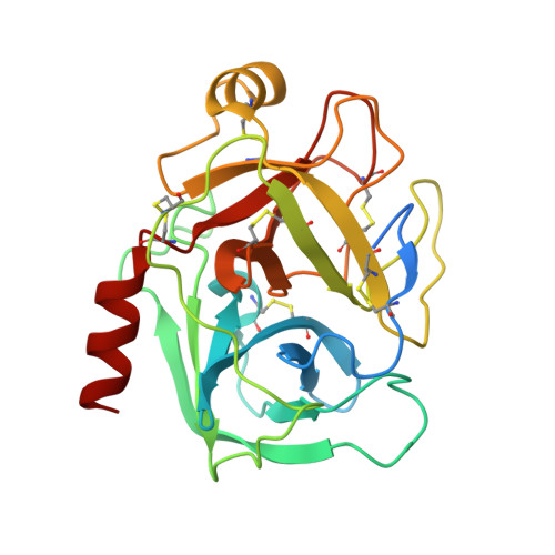
Trypsin fast data collection


Experimental Data Snapshot

  • Method: X-RAY DIFFRACTION
  • Resolution: 1.43 Å
  • R-Value Free: 
    0.206 (Depositor), 0.206 (DCC) 
  • R-Value Work: 
    0.185 (Depositor), 0.185 (DCC) 

Starting Model: experimental
View more details

wwPDB Validation 3D Report Full Report

Validation slider image for 6SWV

This is version 1.2 of the entry. See complete history. 

Literature

ID30A-3 (MASSIF-3) - a beamline for macromolecular crystallography at the ESRF with a small intense beam.

von Stetten, D., Carpentier, P., Flot, D., Beteva, A., Caserotto, H., Dobias, F., Guijarro, M., Giraud, T., Lentini, M., McSweeney, S., Royant, A., Petitdemange, S., Sinoir, J., Surr, J., Svensson, O., Theveneau, P., Leonard, G.A., Mueller-Dieckmann, C.

(2020) J Synchrotron Radiat 27: 844-851

  • DOI: https://doi.org/10.1107/S1600577520004002
  • Primary Citation Related Structures: 
    6SWV, 6Y8G

  • PubMed Abstract: 

    ID30A-3 (or MASSIF-3) is a mini-focus (beam size 18 µm × 14 µm) highly intense (2.0 × 10 13  photons s -1 ), fixed-energy (12.81 keV) beamline for macromolecular crystallography (MX) experiments at the European Synchrotron Radiation Facility (ESRF). MASSIF-3 is one of two fixed-energy beamlines sited on the first branch of the canted undulator setup on the ESRF ID30 port and is equipped with a MD2 micro-diffractometer, a Flex HCD sample changer, and an Eiger X 4M fast hybrid photon-counting detector. MASSIF-3 is recommended for collecting diffraction data from single small crystals (≤15 µm in one dimension) or for experiments using serial methods. The end-station has been in full user operation since December 2014, and here its current characteristics and capabilities are described.


  • Organizational Affiliation: 
    • European Synchrotron Radiation Facility, 71 Avenue des Martyrs, 38000 Grenoble, France.

Macromolecule Content 

  • Total Structure Weight: 26.54 kDa 
  • Atom Count: 1,901 
  • Modeled Residue Count: 223 
  • Deposited Residue Count: 246 
  • Unique protein chains: 1

Macromolecules

Find similar proteins by:|  3D Structure
Entity ID: 1
MoleculeChains  Sequence LengthOrganismDetailsImage
Cationic trypsinA [auth AAA]246Bos taurusMutation(s): 0 
EC: 3.4.21.4
UniProt
Find proteins for P00760 (Bos taurus)
Explore P00760 
Go to UniProtKB:  P00760
Entity Groups
Sequence Clusters30% Identity50% Identity70% Identity90% Identity95% Identity100% Identity
UniProt GroupP00760
Sequence Annotations
Expand
Reference Sequence

Small Molecules

Ligands 3 Unique
IDChains Name / Formula / InChI Key2D Diagram3D Interactions
BEN

Query on BEN


Download:Ideal Coordinates CCD File
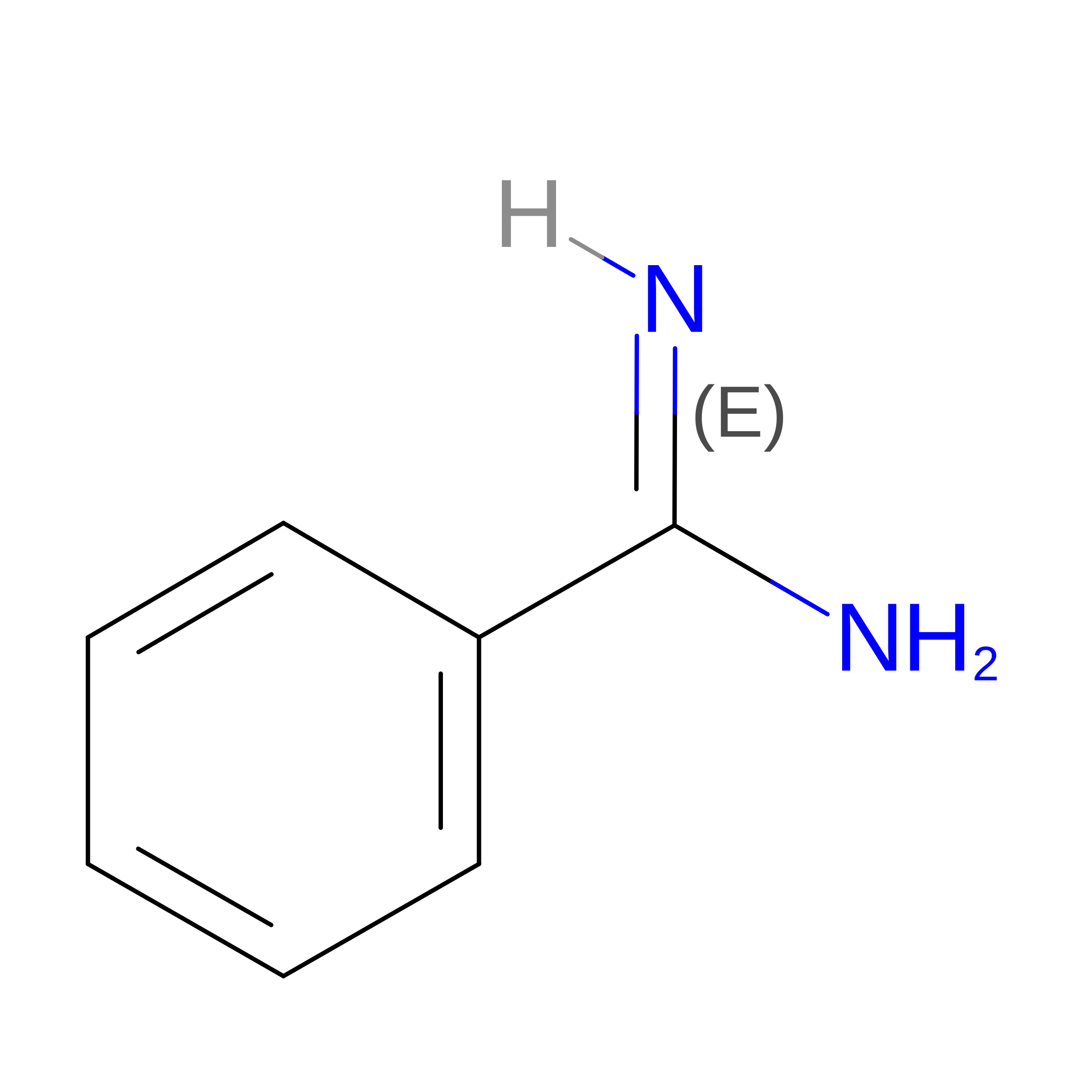
I [auth AAA]BENZAMIDINE
C7 H8 N2
PXXJHWLDUBFPOL-UHFFFAOYSA-N
SO4

Query on SO4


Download:Ideal Coordinates CCD File
C [auth AAA]
D [auth AAA]
E [auth AAA]
F [auth AAA]
G [auth AAA]
C [auth AAA],
D [auth AAA],
E [auth AAA],
F [auth AAA],
G [auth AAA],
H [auth AAA]
SULFATE ION
O4 S
QAOWNCQODCNURD-UHFFFAOYSA-L
CA

Query on CA


Download:Ideal Coordinates CCD File
B [auth AAA]CALCIUM ION
Ca
BHPQYMZQTOCNFJ-UHFFFAOYSA-N

Experimental Data & Validation

Experimental Data

  • Method: X-RAY DIFFRACTION
  • Resolution: 1.43 Å
  • R-Value Free:  0.206 (Depositor), 0.206 (DCC) 
  • R-Value Work:  0.185 (Depositor), 0.185 (DCC) 
Space Group: P 21 21 21
Unit Cell:
Length ( Å )Angle ( ˚ )
a = 54.28α = 90
b = 57.944β = 90
c = 66.947γ = 90
Software Package:
Software NamePurpose
REFMACrefinement
XDSdata reduction
Aimlessdata scaling
PHASERphasing

Structure Validation

View Full Validation Report


Entry History 

Deposition Data

Revision History  (Full details and data files)

  • Version 1.0: 2020-05-20
    Type: Initial release
  • Version 1.1: 2024-01-24
    Changes: Data collection, Database references, Derived calculations, Refinement description
  • Version 1.2: 2024-11-20
    Changes: Structure summary