6STZ | pdb_00006stz

Crystal structure of dimethylated RSLex - cucurbituril free form


Experimental Data Snapshot

  • Method: X-RAY DIFFRACTION
  • Resolution: 1.14 Å
  • R-Value Free: 
    0.198 (Depositor), 0.193 (DCC) 
  • R-Value Work: 
    0.173 (Depositor), 0.174 (DCC) 
  • R-Value Observed: 
    0.174 (Depositor) 

Starting Model: experimental
View more details

wwPDB Validation 3D Report Full Report

Validation slider image for 6STZ

Ligand Structure Quality Assessment 


This is version 1.2 of the entry. See complete history

Literature

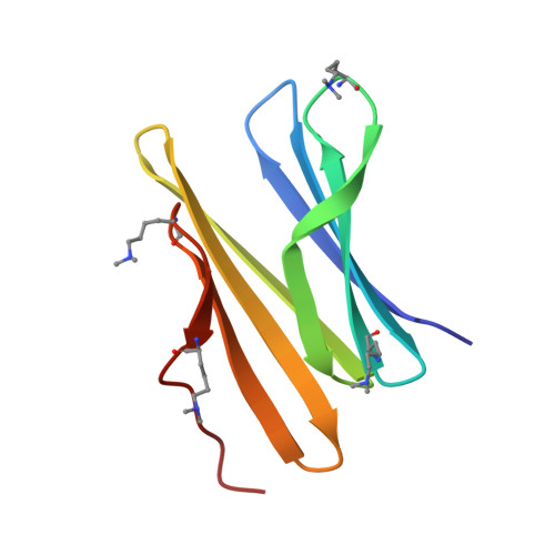
Engineered assembly of a protein-cucurbituril biohybrid.

Guagnini, F.Engilberge, S.Ramberg, K.O.Perez, J.Crowley, P.B.

(2020) Chem Commun (Camb) 56: 360-363

  • DOI: https://doi.org/10.1039/c9cc07198a
  • Primary Citation Related Structures: 
    6STZ, 6SU0

  • PubMed Abstract: 

    A crystalline biohybrid with a 4 : 1 protein : cucurbituril mass ratio is presented. This result was achieved by engineering additional cucurbit[7]uril (Q7) binding sites into a β-propeller protein. In contrast to the parent protein, Q7-controlled assembly of the engineered variant occurred in solution, as evidenced by NMR and SAXS measurements.


  • Organizational Affiliation
    • School of Chemistry, National University of Ireland Galway, University Road, Galway, H91 TK33, Ireland. peter.crowley@nuigalway.ie.

Macromolecule Content 

  • Total Structure Weight: 61.25 kDa 
  • Atom Count: 5,107 
  • Modeled Residue Count: 529 
  • Deposited Residue Count: 534 
  • Unique protein chains: 1

Macromolecules

Find similar proteins by:|  3D Structure
Entity ID: 1
MoleculeChains  Sequence LengthOrganismDetailsImage
Fucose-binding lectin protein
A, B, C, D, E
A, B, C, D, E, F
89Ralstonia solanacearumMutation(s): 1 
Gene Names: rslRUN215_v1_180133
UniProt
Find proteins for Q8XXK6 (Ralstonia nicotianae (strain ATCC BAA-1114 / GMI1000))
Explore Q8XXK6 
Go to UniProtKB:  Q8XXK6
Entity Groups
Sequence Clusters30% Identity50% Identity70% Identity90% Identity95% Identity100% Identity
UniProt GroupQ8XXK6
Sequence Annotations
Expand
Reference Sequence

Small Molecules

Ligands 1 Unique
IDChains Name / Formula / InChI Key2D Diagram3D Interactions
BDF

Query on BDF


Download:Ideal Coordinates CCD File
G [auth A]
H [auth A]
I [auth B]
J [auth B]
K [auth C]
G [auth A],
H [auth A],
I [auth B],
J [auth B],
K [auth C],
L [auth C],
M [auth D],
N [auth D],
O [auth E],
P [auth E],
Q [auth F],
R [auth F]
beta-D-fructopyranose
C6 H12 O6
LKDRXBCSQODPBY-ARQDHWQXSA-N
Modified Residues  1 Unique
IDChains TypeFormula2D DiagramParent
MLY
Query on MLY
A, B, C, D, E
A, B, C, D, E, F
L-PEPTIDE LINKINGC8 H18 N2 O2LYS

Experimental Data & Validation

Experimental Data

  • Method: X-RAY DIFFRACTION
  • Resolution: 1.14 Å
  • R-Value Free:  0.198 (Depositor), 0.193 (DCC) 
  • R-Value Work:  0.173 (Depositor), 0.174 (DCC) 
  • R-Value Observed: 0.174 (Depositor) 
Space Group: P 32
Unit Cell:
Length ( Å )Angle ( ˚ )
a = 43.948α = 90
b = 43.948β = 90
c = 207.273γ = 120
Software Package:
Software NamePurpose
BUSTERrefinement
Aimlessdata scaling
PDB_EXTRACTdata extraction
XDSdata reduction
PHASERphasing

Structure Validation

View Full Validation Report


Ligand Structure Quality Assessment 


Entry History 

& Funding Information

Deposition Data


Funding OrganizationLocationGrant Number
Science Foundation IrelandIreland13/CDA/2168

Revision History  (Full details and data files)

  • Version 1.0: 2020-02-19
    Type: Initial release
  • Version 1.1: 2020-07-29
    Type: Remediation
    Reason: Carbohydrate remediation
    Changes: Data collection, Derived calculations, Structure summary
  • Version 1.2: 2024-01-24
    Changes: Data collection, Database references, Refinement description, Structure summary