6SMT | pdb_00006smt

S-enantioselective imine reductase from Mycobacterium smegmatis


Experimental Data Snapshot

  • Method: X-RAY DIFFRACTION
  • Resolution: 1.55 Å
  • R-Value Free: 
    0.155 (Depositor), 0.155 (DCC) 
  • R-Value Work: 
    0.131 (Depositor), 0.131 (DCC) 
  • R-Value Observed: 
    0.132 (Depositor) 

Starting Models: experimental
View more details

wwPDB Validation 3D Report Full Report

Validation slider image for 6SMT

Ligand Structure Quality Assessment 


This is version 1.3 of the entry. See complete history

Literature

Structural Characterization of an S -enantioselective Imine Reductase from Mycobacterium Smegmatis .

Meyer, T.Zumbragel, N.Geerds, C.Groger, H.Niemann, H.H.

(2020) Biomolecules 10

  • DOI: https://doi.org/10.3390/biom10081130
  • Primary Citation Related Structures: 
    6SMT

  • PubMed Abstract: 

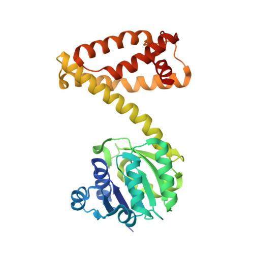
    NADPH-dependent imine reductases (IREDs) are enzymes capable of enantioselectively reducing imines to chiral secondary amines, which represent important building blocks in the chemical and pharmaceutical industry. Since their discovery in 2011, many previously unknown IREDs have been identified, biochemically and structurally characterized and categorized into families. However, the catalytic mechanism and guiding principles for substrate specificity and stereoselectivity remain disputed. Herein, we describe the crystal structure of S -IRED- Ms from Mycobacterium smegmatis together with its cofactor NADPH. S -IRED- Ms belongs to the S -enantioselective superfamily 3 (SFam3) and is the first IRED from SFam3 to be structurally described. The data presented provide further evidence for the overall high degree of structural conservation between different IREDs of various superfamilies. We discuss the role of Asp170 in catalysis and the importance of hydrophobic amino acids in the active site for stereospecificity. Moreover, a separate entrance to the active site, potentially functioning according to a gatekeeping mechanism regulating access and, therefore, substrate specificity is described.


  • Organizational Affiliation
    • Structural Biochemistry, Faculty of Chemistry, Bielefeld University, Universitätsstraße 25, 33615 Bielefeld, Germany.

Macromolecule Content 

  • Total Structure Weight: 160.67 kDa 
  • Atom Count: 13,314 
  • Modeled Residue Count: 1,432 
  • Deposited Residue Count: 1,490 
  • Unique protein chains: 1

Macromolecules

Find similar proteins by:|  3D Structure
Entity ID: 1
MoleculeChains  Sequence LengthOrganismDetailsImage
6-phosphogluconate dehydrogenase, NAD-binding
A, B, C, D, E
298Mycolicibacterium smegmatis MC2 155Mutation(s): 0 
Gene Names: MSMEG_6341
UniProt
Find proteins for A0R5X0 (Mycolicibacterium smegmatis (strain ATCC 700084 / mc(2)155))
Explore A0R5X0 
Go to UniProtKB:  A0R5X0
Entity Groups
Sequence Clusters30% Identity50% Identity70% Identity90% Identity95% Identity100% Identity
UniProt GroupA0R5X0
Sequence Annotations
Expand
Reference Sequence

Small Molecules

Ligands 6 Unique
IDChains Name / Formula / InChI Key2D Diagram3D Interactions
NDP

Query on NDP


Download:Ideal Coordinates CCD File
AA [auth E],
F [auth A],
L [auth B],
P [auth C],
T [auth D]
NADPH DIHYDRO-NICOTINAMIDE-ADENINE-DINUCLEOTIDE PHOSPHATE
C21 H30 N7 O17 P3
ACFIXJIJDZMPPO-NNYOXOHSSA-N
2EH

Query on 2EH


Download:Ideal Coordinates CCD File
HA [auth E],
J [auth A],
N [auth B],
S [auth C],
Y [auth D]
(2S)-2-ethylhexan-1-ol
C8 H18 O
YIWUKEYIRIRTPP-QMMMGPOBSA-N
PEG

Query on PEG


Download:Ideal Coordinates CCD File
BA [auth E]DI(HYDROXYETHYL)ETHER
C4 H10 O3
MTHSVFCYNBDYFN-UHFFFAOYSA-N
GOL

Query on GOL


Download:Ideal Coordinates CCD File
K [auth A],
O [auth B],
Z [auth D]
GLYCEROL
C3 H8 O3
PEDCQBHIVMGVHV-UHFFFAOYSA-N
EDO

Query on EDO


Download:Ideal Coordinates CCD File
GA [auth E],
I [auth A],
R [auth C]
1,2-ETHANEDIOL
C2 H6 O2
LYCAIKOWRPUZTN-UHFFFAOYSA-N
FMT

Query on FMT


Download:Ideal Coordinates CCD File
CA [auth E]
DA [auth E]
EA [auth E]
FA [auth E]
G [auth A]
CA [auth E],
DA [auth E],
EA [auth E],
FA [auth E],
G [auth A],
H [auth A],
M [auth B],
Q [auth C],
U [auth D],
V [auth D],
W [auth D],
X [auth D]
FORMIC ACID
C H2 O2
BDAGIHXWWSANSR-UHFFFAOYSA-N

Experimental Data & Validation

Experimental Data

  • Method: X-RAY DIFFRACTION
  • Resolution: 1.55 Å
  • R-Value Free:  0.155 (Depositor), 0.155 (DCC) 
  • R-Value Work:  0.131 (Depositor), 0.131 (DCC) 
  • R-Value Observed: 0.132 (Depositor) 
Space Group: I 1 2 1
Unit Cell:
Length ( Å )Angle ( ˚ )
a = 117.007α = 90
b = 77.023β = 90.2
c = 194.485γ = 90
Software Package:
Software NamePurpose
REFMACrefinement
XDSdata reduction
Aimlessdata scaling
PHASERphasing
PDB_EXTRACTdata extraction

Structure Validation

View Full Validation Report


Ligand Structure Quality Assessment 


Entry History 

& Funding Information

Deposition Data


Funding OrganizationLocationGrant Number
German Federal Ministry for Education and ResearchGermany031A184A

Revision History  (Full details and data files)

  • Version 1.0: 2020-08-12
    Type: Initial release
  • Version 1.1: 2020-08-19
    Changes: Database references
  • Version 1.2: 2020-09-30
    Changes: Database references
  • Version 1.3: 2024-01-24
    Changes: Data collection, Database references, Refinement description