6SLN | pdb_00006sln

Structure of the RagAB peptide transporter


Experimental Data Snapshot

  • Method: X-RAY DIFFRACTION
  • Resolution: 2.61 Å
  • R-Value Free: 
    0.264 (Depositor), 0.264 (DCC) 
  • R-Value Work: 
    0.207 (Depositor), 0.208 (DCC) 
  • R-Value Observed: 
    0.210 (Depositor) 

Starting Model: experimental
View more details

wwPDB Validation 3D Report Full Report

Validation slider image for 6SLN

Ligand Structure Quality Assessment 


This is version 1.3 of the entry. See complete history

Literature

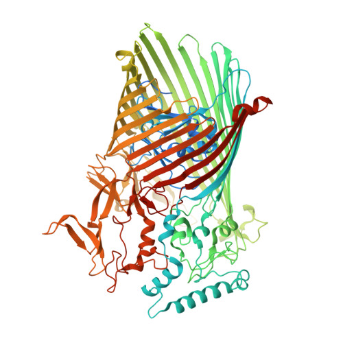
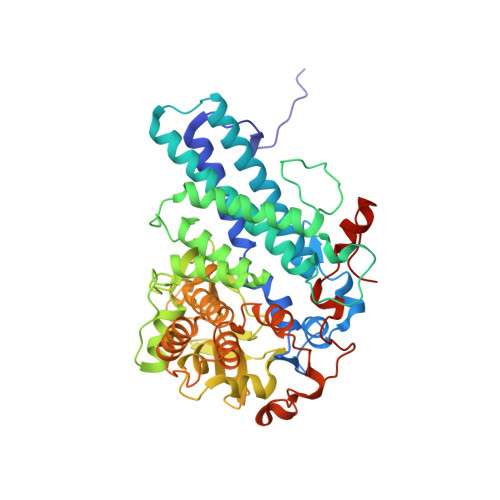
Structural and functional insights into oligopeptide acquisition by the RagAB transporter from Porphyromonas gingivalis.

Madej, M.White, J.B.R.Nowakowska, Z.Rawson, S.Scavenius, C.Enghild, J.J.Bereta, G.P.Pothula, K.Kleinekathoefer, U.Basle, A.Ranson, N.A.Potempa, J.van den Berg, B.

(2020) Nat Microbiol 5: 1016-1025

  • DOI: https://doi.org/10.1038/s41564-020-0716-y
  • Primary Citation Related Structures: 
    6SLI, 6SLJ, 6SLN, 6SM3, 6SML, 6SMQ

  • PubMed Abstract: 

    Porphyromonas gingivalis, an asaccharolytic member of the Bacteroidetes, is a keystone pathogen in human periodontitis that may also contribute to the development of other chronic inflammatory diseases. P. gingivalis utilizes protease-generated peptides derived from extracellular proteins for growth, but how these peptides enter the cell is not clear. Here, we identify RagAB as the outer-membrane importer for these peptides. X-ray crystal structures show that the transporter forms a dimeric RagA 2 B 2 complex, with the RagB substrate-binding surface-anchored lipoprotein forming a closed lid on the RagA TonB-dependent transporter. Cryo-electron microscopy structures reveal the opening of the RagB lid and thus provide direct evidence for a 'pedal bin' mechanism of nutrient uptake. Together with mutagenesis, peptide-binding studies and RagAB peptidomics, our work identifies RagAB as a dynamic, selective outer-membrane oligopeptide-acquisition machine that is essential for the efficient utilization of proteinaceous nutrients by P. gingivalis.


  • Organizational Affiliation
    • Department of Microbiology, Faculty of Biochemistry, Biophysics and Biotechnology, Jagiellonian University, Krakow, Poland.

Macromolecule Content 

  • Total Structure Weight: 338.23 kDa 
  • Atom Count: 22,773 
  • Modeled Residue Count: 2,794 
  • Deposited Residue Count: 2,996 
  • Unique protein chains: 3

Macromolecules

Find similar proteins by:|  3D Structure
Entity ID: 1
MoleculeChains  Sequence LengthOrganismDetailsImage
RagA protein
A, B
997Porphyromonas gingivalis W83Mutation(s): 0 
Membrane Entity: Yes 
UniProt
Find proteins for Q7MXJ7 (Porphyromonas gingivalis (strain ATCC BAA-308 / W83))
Explore Q7MXJ7 
Go to UniProtKB:  Q7MXJ7
Entity Groups
Sequence Clusters30% Identity50% Identity70% Identity90% Identity95% Identity100% Identity
UniProt GroupQ7MXJ7
Sequence Annotations
Expand
Reference Sequence
Find similar proteins by:|  3D Structure
Entity ID: 2
MoleculeChains  Sequence LengthOrganismDetailsImage
Lipoprotein RagB
C, D
488Porphyromonas gingivalis W83Mutation(s): 0 
Membrane Entity: Yes 
UniProt
Find proteins for F5H948 (Porphyromonas gingivalis (strain ATCC BAA-308 / W83))
Explore F5H948 
Go to UniProtKB:  F5H948
Entity Groups
Sequence Clusters30% Identity50% Identity70% Identity90% Identity95% Identity100% Identity
UniProt GroupF5H948
Sequence Annotations
Expand
Reference Sequence
Find similar proteins by:  Sequence   |   3D Structure  
Entity ID: 3
MoleculeChains  Sequence LengthOrganismDetailsImage
GLN-THR-ALA-GLY-ALA-ASN-SER-GLN-ARG-GLY-SER-ALA-GLYE [auth P],
F [auth Q]
13Porphyromonas gingivalis W83Mutation(s): 0 

Small Molecules

Ligands 4 Unique
IDChains Name / Formula / InChI Key2D Diagram3D Interactions
3PE

Query on 3PE


Download:Ideal Coordinates CCD File
G [auth A],
K [auth B]
1,2-Distearoyl-sn-glycerophosphoethanolamine
C41 H82 N O8 P
LVNGJLRDBYCPGB-LDLOPFEMSA-N
LMT

Query on LMT


Download:Ideal Coordinates CCD File
H [auth A],
L [auth B],
O [auth C],
Q [auth D]
DODECYL-BETA-D-MALTOSIDE
C24 H46 O11
NLEBIOOXCVAHBD-QKMCSOCLSA-N
C8E

Query on C8E


Download:Ideal Coordinates CCD File
I [auth A],
J [auth A],
M [auth B]
(HYDROXYETHYLOXY)TRI(ETHYLOXY)OCTANE
C16 H34 O5
FEOZZFHAVXYAMB-UHFFFAOYSA-N
PLM

Query on PLM


Download:Ideal Coordinates CCD File
N [auth C],
P [auth D]
PALMITIC ACID
C16 H32 O2
IPCSVZSSVZVIGE-UHFFFAOYSA-N

Experimental Data & Validation

Experimental Data

  • Method: X-RAY DIFFRACTION
  • Resolution: 2.61 Å
  • R-Value Free:  0.264 (Depositor), 0.264 (DCC) 
  • R-Value Work:  0.207 (Depositor), 0.208 (DCC) 
  • R-Value Observed: 0.210 (Depositor) 
Space Group: P 21 21 21
Unit Cell:
Length ( Å )Angle ( ˚ )
a = 129.781α = 90
b = 142.734β = 90
c = 250.048γ = 90
Software Package:
Software NamePurpose
PHENIXrefinement
PHENIXrefinement
DIALSdata reduction
Aimlessdata scaling
PHASERphasing

Structure Validation

View Full Validation Report


Ligand Structure Quality Assessment 


Entry History 

Deposition Data

Revision History  (Full details and data files)

  • Version 1.0: 2020-05-20
    Type: Initial release
  • Version 1.1: 2020-08-05
    Changes: Database references, Derived calculations
  • Version 1.2: 2024-01-24
    Changes: Data collection, Database references, Derived calculations, Refinement description, Structure summary
  • Version 1.3: 2025-10-01
    Changes: Advisory, Derived calculations, Structure summary