6SHS | pdb_00006shs

Abeta fibril (Morphology I)


Experimental Data Snapshot

  • Method: ELECTRON MICROSCOPY
  • Resolution: 4.40 Å
  • Aggregation State: HELICAL ARRAY 
  • Reconstruction Method: SINGLE PARTICLE 

wwPDB Validation 3D Report Full Report

Validation slider image for 6SHS

This is version 1.1 of the entry. See complete history

Literature

Cryo-EM structure and polymorphism of A beta amyloid fibrils purified from Alzheimer's brain tissue.

Kollmer, M.Close, W.Funk, L.Rasmussen, J.Bsoul, A.Schierhorn, A.Schmidt, M.Sigurdson, C.J.Jucker, M.Fandrich, M.

(2019) Nat Commun 10: 4760-4760

  • DOI: https://doi.org/10.1038/s41467-019-12683-8
  • Primary Citation Related Structures: 
    6SHS

  • PubMed Abstract: 

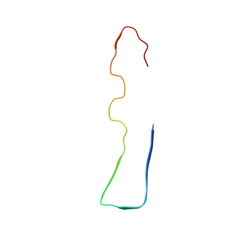
    The formation of Aβ amyloid fibrils is a neuropathological hallmark of Alzheimer's disease and cerebral amyloid angiopathy. However, the structure of Aβ amyloid fibrils from brain tissue is poorly understood. Here we report the purification of Aβ amyloid fibrils from meningeal Alzheimer's brain tissue and their structural analysis with cryo-electron microscopy. We show that these fibrils are polymorphic but consist of similarly structured protofilaments. Brain derived Aβ amyloid fibrils are right-hand twisted and their peptide fold differs sharply from previously analyzed Aβ fibrils that were formed in vitro. These data underscore the importance to use patient-derived amyloid fibrils when investigating the structural basis of the disease.


  • Organizational Affiliation
    • Institute of Protein Biochemistry, Ulm University, Helmholtzstrasse 8/1, 89081, Ulm, Germany.

Macromolecule Content 

  • Total Structure Weight: 52.03 kDa 
  • Atom Count: 3,660 
  • Modeled Residue Count: 480 
  • Deposited Residue Count: 480 
  • Unique protein chains: 1

Macromolecules

Find similar proteins by:|  3D Structure
Entity ID: 1
MoleculeChains  Sequence LengthOrganismDetailsImage
Amyloid-beta precursor protein40Homo sapiensMutation(s): 0 
UniProt & NIH Common Fund Data Resources
Find proteins for P05067 (Homo sapiens)
Explore P05067 
Go to UniProtKB:  P05067
PHAROS:  P05067
GTEx:  ENSG00000142192 
Entity Groups
Sequence Clusters30% Identity50% Identity70% Identity90% Identity95% Identity100% Identity
UniProt GroupP05067
Sequence Annotations
Expand
Reference Sequence

Experimental Data & Validation

Experimental Data

  • Method: ELECTRON MICROSCOPY
  • Resolution: 4.40 Å
  • Aggregation State: HELICAL ARRAY 
  • Reconstruction Method: SINGLE PARTICLE 
EM Software:
TaskSoftware PackageVersion
RECONSTRUCTIONRELION
MODEL REFINEMENTPHENIX

Structure Validation

View Full Validation Report


Entry History 

& Funding Information

Deposition Data


Funding OrganizationLocationGrant Number
German Research FoundationGermanyFA456/12-1
German Research FoundationGermanyFA456/24-1

Revision History  (Full details and data files)

  • Version 1.0: 2019-11-06
    Type: Initial release
  • Version 1.1: 2024-05-22
    Changes: Data collection, Database references