6SCD | pdb_00006scd

Polyester hydrolase PE-H Y250S mutant of Pseudomonas aestusnigri


Experimental Data Snapshot

  • Method: X-RAY DIFFRACTION
  • Resolution: 1.35 Å
  • R-Value Free: 
    0.152 (Depositor), 0.171 (DCC) 
  • R-Value Work: 
    0.113 (Depositor) 
  • R-Value Observed: 
    0.115 (Depositor) 

Starting Model: experimental
View more details

wwPDB Validation 3D Report Full Report

Validation slider image for 6SCD

This is version 1.3 of the entry. See complete history

Literature

A Novel Polyester Hydrolase From the Marine BacteriumPseudomonas aestusnigri -Structural and Functional Insights.

Bollinger, A.Thies, S.Knieps-Grunhagen, E.Gertzen, C.Kobus, S.Hoppner, A.Ferrer, M.Gohlke, H.Smits, S.H.J.Jaeger, K.E.

(2020) Front Microbiol 11: 114-114

  • DOI: https://doi.org/10.3389/fmicb.2020.00114
  • Primary Citation Related Structures: 
    6SBN, 6SCD

  • PubMed Abstract: 

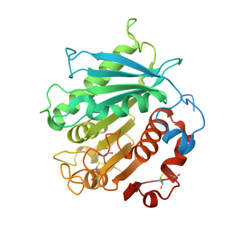
    Biodegradation of synthetic polymers, in particular polyethylene terephthalate (PET), is of great importance, since environmental pollution with PET and other plastics has become a severe global problem. Here, we report on the polyester degrading ability of a novel carboxylic ester hydrolase identified in the genome of the marine hydrocarbonoclastic bacterium Pseudomonas aestusnigri VGXO14 T . The enzyme, designated PE-H, belongs to the type IIa family of PET hydrolytic enzymes as indicated by amino acid sequence homology. It was produced in Escherichia coli , purified and its crystal structure was solved at 1.09 Å resolution representing the first structure of a type IIa PET hydrolytic enzyme. The structure shows a typical α/β-hydrolase fold and high structural homology to known polyester hydrolases. PET hydrolysis was detected at 30°C with amorphous PET film (PETa), but not with PET film from a commercial PET bottle (PETb). A rational mutagenesis study to improve the PET degrading potential of PE-H yielded variant PE-H (Y250S) which showed improved activity, ultimately also allowing the hydrolysis of PETb. The crystal structure of this variant solved at 1.35 Å resolution allowed to rationalize the improvement of enzymatic activity. A PET oligomer binding model was proposed by molecular docking computations. Our results indicate a significant potential of the marine bacterium P. aestusnigri for PET degradation.


  • Organizational Affiliation
    • Institute of Molecular Enzyme Technology, Heinrich Heine University Düsseldorf, Jülich, Germany.

Macromolecule Content 

  • Total Structure Weight: 67.56 kDa 
  • Atom Count: 4,590 
  • Modeled Residue Count: 532 
  • Deposited Residue Count: 624 
  • Unique protein chains: 1

Macromolecules

Find similar proteins by:|  3D Structure
Entity ID: 1
MoleculeChains  Sequence LengthOrganismDetailsImage
Polyester hydrolase
A, B
312Halopseudomonas aestusnigriMutation(s): 1 
Gene Names: B7O88_11480

Small Molecules

Ligands 7 Unique
IDChains Name / Formula / InChI Key2D Diagram3D Interactions
PEG

Query on PEG


Download:Ideal Coordinates CCD File
I [auth A]DI(HYDROXYETHYL)ETHER
C4 H10 O3
MTHSVFCYNBDYFN-UHFFFAOYSA-N
SO4

Query on SO4


Download:Ideal Coordinates CCD File
C [auth A]SULFATE ION
O4 S
QAOWNCQODCNURD-UHFFFAOYSA-L
PO4

Query on PO4


Download:Ideal Coordinates CCD File
D [auth A],
E [auth A],
M [auth B]
PHOSPHATE ION
O4 P
NBIIXXVUZAFLBC-UHFFFAOYSA-K
GOL

Query on GOL


Download:Ideal Coordinates CCD File
J [auth A],
K [auth A]
GLYCEROL
C3 H8 O3
PEDCQBHIVMGVHV-UHFFFAOYSA-N
ACT

Query on ACT


Download:Ideal Coordinates CCD File
L [auth A]ACETATE ION
C2 H3 O2
QTBSBXVTEAMEQO-UHFFFAOYSA-M
CL

Query on CL


Download:Ideal Coordinates CCD File
G [auth A],
H [auth A],
P [auth B]
CHLORIDE ION
Cl
VEXZGXHMUGYJMC-UHFFFAOYSA-M
NA

Query on NA


Download:Ideal Coordinates CCD File
F [auth A],
N [auth B],
O [auth B]
SODIUM ION
Na
FKNQFGJONOIPTF-UHFFFAOYSA-N

Experimental Data & Validation

Experimental Data

  • Method: X-RAY DIFFRACTION
  • Resolution: 1.35 Å
  • R-Value Free:  0.152 (Depositor), 0.171 (DCC) 
  • R-Value Work:  0.113 (Depositor) 
  • R-Value Observed: 0.115 (Depositor) 
Space Group: I 21 21 21
Unit Cell:
Length ( Å )Angle ( ˚ )
a = 94.526α = 90
b = 98.268β = 90
c = 121.233γ = 90
Software Package:
Software NamePurpose
REFMACrefinement
PDB_EXTRACTdata extraction
XDSdata reduction
XDSdata scaling
PHASERphasing

Structure Validation

View Full Validation Report


Entry History 

Deposition Data

Revision History  (Full details and data files)

  • Version 1.0: 2020-02-26
    Type: Initial release
  • Version 1.1: 2020-03-11
    Changes: Database references
  • Version 1.2: 2024-01-24
    Changes: Data collection, Database references, Derived calculations, Refinement description
  • Version 1.3: 2024-11-20
    Changes: Structure summary