6SAQ

wild-type NuoEF from Aquifex aeolicus bound to NADH-OH


Experimental Data Snapshot

  • Method: X-RAY DIFFRACTION
  • Resolution: 2.02 Å
  • R-Value Free: 0.181 
  • R-Value Work: 0.159 
  • R-Value Observed: 0.160 

Starting Model: experimental
View more details

wwPDB Validation   3D Report Full Report


Ligand Structure Quality Assessment 


This is version 1.3 of the entry. See complete history


Literature

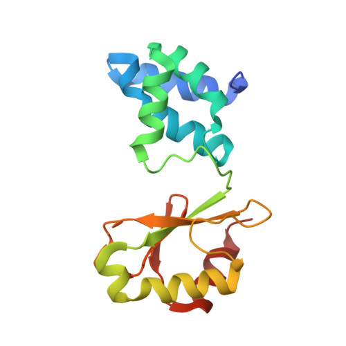
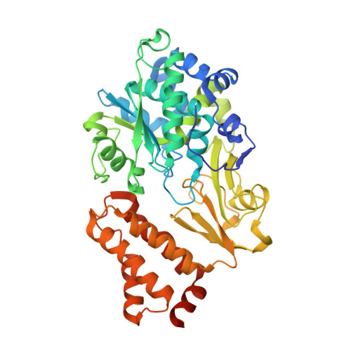
Structural Basis for Inhibition of ROS-Producing Respiratory Complex I by NADH-OH.

Friedrich, T.Vranas, M.Wohlwend, D.Qiu, D.Gerhardt, S.Trncik, C.Pervaiz, M.Ritter, K.Steimle, S.Randazzo, A.Einsle, O.Gunther, S.Jessen, H.J.Kotlyar, A.

(2021) Angew Chem Int Ed Engl 

  • DOI: https://doi.org/10.1002/anie.202112165
  • Primary Citation of Related Structures:  
    6SAQ

  • PubMed Abstract: 

    NADH:ubiquinone oxidoreductase, respiratory complex I, plays a central role in cellular energy metabolism. As a major source of reactive oxygen species (ROS) it affects ageing and mitochondrial dysfunction. The novel inhibitor NADH-OH specifically blocks NADH oxidation and ROS production by complex I in nanomolar concentrations. Attempts to elucidate its structure by NMR spectroscopy have failed. Here, by using X-ray crystallographic analysis, we report the structure of NADH-OH bound in the active site of a soluble fragment of complex I at 2.0 Å resolution. We have identified key amino acid residues that are specific and essential for binding NADH-OH. Furthermore, the structure sheds light on the specificity of NADH-OH towards the unique Rossmann-fold of complex I and indicates a regulatory role in mitochondrial ROS generation. In addition, NADH-OH acts as a lead-structure for the synthesis of a novel class of ROS suppressors.


  • Organizational Affiliation

    Institute of Biochemistry, University of Freiburg, 79104, Freiburg, Germany.


Macromolecules
Find similar proteins by:  (by identity cutoff)  |  3D Structure
Entity ID: 1
MoleculeChains Sequence LengthOrganismDetailsImage
NADH-quinone oxidoreductase subunit E
A, C
160Aquifex aeolicus VF5Mutation(s): 0 
Gene Names: nuoEaq_574
EC: 7.1.1
UniProt
Find proteins for O66842 (Aquifex aeolicus (strain VF5))
Explore O66842 
Go to UniProtKB:  O66842
Entity Groups  
Sequence Clusters30% Identity50% Identity70% Identity90% Identity95% Identity100% Identity
UniProt GroupO66842
Sequence Annotations
Expand
  • Reference Sequence
Find similar proteins by:  (by identity cutoff)  |  3D Structure
Entity ID: 2
MoleculeChains Sequence LengthOrganismDetailsImage
NADH-quinone oxidoreductase subunit F
B, D
434Aquifex aeolicus VF5Mutation(s): 0 
Gene Names: nuoFaq_573
EC: 7.1.1
UniProt
Find proteins for O66841 (Aquifex aeolicus (strain VF5))
Explore O66841 
Go to UniProtKB:  O66841
Entity Groups  
Sequence Clusters30% Identity50% Identity70% Identity90% Identity95% Identity100% Identity
UniProt GroupO66841
Sequence Annotations
Expand
  • Reference Sequence
Small Molecules
Ligands 5 Unique
IDChains Name / Formula / InChI Key2D Diagram3D Interactions
L3W (Subject of Investigation/LOI)
Query on L3W

Download Ideal Coordinates CCD File 
J [auth B],
Q [auth D]
[[(2~{R},3~{S},4~{R},5~{R})-5-(6-aminopurin-9-yl)-3,4-bis(oxidanyl)oxolan-2-yl]methoxy-oxidanyl-phosphoryl] [(2~{R},3~{S},4~{R},5~{R})-5-[[(1~{E},3~{Z})-5-azanyl-4-oxidanyl-5-oxidanylidene-penta-1,3-dienyl]-methanoyl-amino]-3,4-bis(oxidanyl)oxolan-2-yl]methyl hydrogen phosphate
C21 H29 N7 O16 P2
QSTZUQWNOOUKGF-OTTADCDWSA-N
FMN
Query on FMN

Download Ideal Coordinates CCD File 
H [auth B],
O [auth D]
FLAVIN MONONUCLEOTIDE
C17 H21 N4 O9 P
FVTCRASFADXXNN-SCRDCRAPSA-N
SF4
Query on SF4

Download Ideal Coordinates CCD File 
I [auth B],
P [auth D]
IRON/SULFUR CLUSTER
Fe4 S4
LJBDFODJNLIPKO-UHFFFAOYSA-N
FES
Query on FES

Download Ideal Coordinates CCD File 
E [auth A],
N [auth C]
FE2/S2 (INORGANIC) CLUSTER
Fe2 S2
NIXDOXVAJZFRNF-UHFFFAOYSA-N
GOL
Query on GOL

Download Ideal Coordinates CCD File 
F [auth A]
G [auth A]
K [auth B]
L [auth B]
M [auth B]
F [auth A],
G [auth A],
K [auth B],
L [auth B],
M [auth B],
R [auth D],
S [auth D]
GLYCEROL
C3 H8 O3
PEDCQBHIVMGVHV-UHFFFAOYSA-N
Experimental Data & Validation

Experimental Data

  • Method: X-RAY DIFFRACTION
  • Resolution: 2.02 Å
  • R-Value Free: 0.181 
  • R-Value Work: 0.159 
  • R-Value Observed: 0.160 
  • Space Group: P 1 21 1
Unit Cell:
Length ( Å )Angle ( ˚ )
a = 95.976α = 90
b = 63.794β = 105.65
c = 121.398γ = 90
Software Package:
Software NamePurpose
BUSTERrefinement
XDSdata reduction
autoPROCdata scaling
MOLREPphasing

Structure Validation

View Full Validation Report


Ligand Structure Quality Assessment 


Entry History & Funding Information

Deposition Data

  • Released Date: 2020-07-22 
  • Deposition Author(s): Gerhardt, S.

Funding OrganizationLocationGrant Number
German Research FoundationGermany-278002225/RTG 2202
German Research FoundationGermany-235777276/RTG 1976
German Research FoundationGermanySPP 1927

Revision History  (Full details and data files)

  • Version 1.0: 2020-07-22
    Type: Initial release
  • Version 1.1: 2020-07-29
    Changes: Derived calculations
  • Version 1.2: 2021-10-13
    Changes: Data collection, Database references
  • Version 1.3: 2024-01-24
    Changes: Data collection, Refinement description