6S84 | pdb_00006s84

TsaBDE complex from Thermotoga maritima


Experimental Data Snapshot

  • Method: X-RAY DIFFRACTION
  • Resolution: 2.89 Å
  • R-Value Free: 
    0.282 (Depositor), 0.271 (DCC) 
  • R-Value Work: 
    0.209 (Depositor), 0.199 (DCC) 
  • R-Value Observed: 
    0.212 (Depositor) 

wwPDB Validation 3D Report Full Report

Validation slider image for 6S84

Ligand Structure Quality Assessment 


This is version 1.1 of the entry. See complete history

Literature

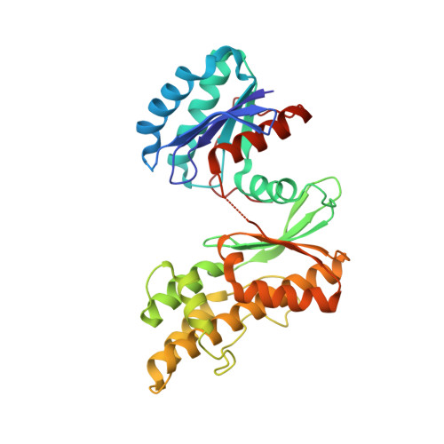
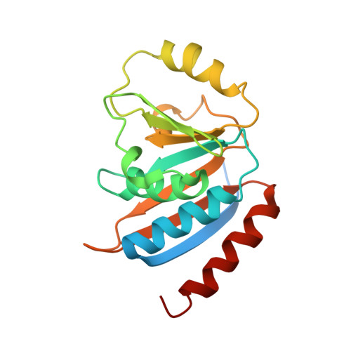
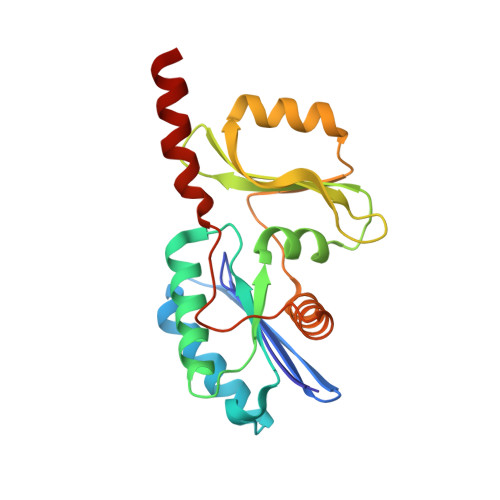
The structure of the TsaB/TsaD/TsaE complex reveals an unexpected mechanism for the bacterial t6A tRNA-modification.

Missoury, S.Plancqueel, S.Li de la Sierra-Gallay, I.Zhang, W.Liger, D.Durand, D.Dammak, R.Collinet, B.van Tilbeurgh, H.

(2018) Nucleic Acids Res 46: 5850-5860

  • DOI: https://doi.org/10.1093/nar/gky323
  • Primary Citation Related Structures: 
    6S84

  • PubMed Abstract: 

    The universal N6-threonylcarbamoyladenosine (t6A) modification at position A37 of ANN-decoding tRNAs is essential for translational fidelity. In bacteria the TsaC enzyme first synthesizes an l-threonylcarbamoyladenylate (TC-AMP) intermediate. In cooperation with TsaB and TsaE, TsaD then transfers the l-threonylcarbamoyl-moiety from TC-AMP onto tRNA. We determined the crystal structure of the TsaB-TsaE-TsaD (TsaBDE) complex of Thermotoga maritima in presence of a non-hydrolysable AMPCPP. TsaE is positioned at the entrance of the active site pocket of TsaD, contacting both the TsaB and TsaD subunits and prohibiting simultaneous tRNA binding. AMPCPP occupies the ATP binding site of TsaE and is sandwiched between TsaE and TsaD. Unexpectedly, the binding of TsaE partially denatures the active site of TsaD causing loss of its essential metal binding sites. TsaE interferes in a pre- or post-catalytic step and its binding to TsaBD is regulated by ATP hydrolysis. This novel binding mode and activation mechanism of TsaE offers good opportunities for antimicrobial drug development.


  • Organizational Affiliation
    • Institute for Integrative Biology of the Cell (I2BC), CEA, CNRS UMR 9198, Univ. Paris-Sud, Université Paris-Saclay, 91198 Gif sur Yvette Cedex, France.

Macromolecule Content 

  • Total Structure Weight: 162.61 kDa 
  • Atom Count: 10,687 
  • Modeled Residue Count: 1,341 
  • Deposited Residue Count: 1,434 
  • Unique protein chains: 3

Macromolecules

Find similar proteins by:|  3D Structure
Entity ID: 1
MoleculeChains  Sequence LengthOrganismDetailsImage
tRNA N6-adenosine threonylcarbamoyltransferaseA,
C [auth D]
327Thermotoga maritima MSB8Mutation(s): 0 
Gene Names: tsaDgcpTM_0145
EC: 2.3.1.234
UniProt
Find proteins for Q9WXZ2 (Thermotoga maritima (strain ATCC 43589 / DSM 3109 / JCM 10099 / NBRC 100826 / MSB8))
Explore Q9WXZ2 
Go to UniProtKB:  Q9WXZ2
Entity Groups
Sequence Clusters30% Identity50% Identity70% Identity90% Identity95% Identity100% Identity
UniProt GroupQ9WXZ2
Sequence Annotations
Expand
Reference Sequence
Find similar proteins by:|  3D Structure
Entity ID: 2
MoleculeChains  Sequence LengthOrganismDetailsImage
ATPase YjeE, predicted to have essential role in cell wall biosynthesisB [auth E],
F [auth B]
184Thermotoga maritima MSB8Mutation(s): 0 
Gene Names: Tmari_1641
Find similar proteins by:|  3D Structure
Entity ID: 3
MoleculeChains  Sequence LengthOrganismDetailsImage
tRNA threonylcarbamoyladenosine biosynthesis protein TsaBD [auth C],
E [auth F]
206Thermotoga maritima MSB8Mutation(s): 0 
Gene Names: tsaBTM_0874Tmari_0876
UniProt
Find proteins for Q9WZX7 (Thermotoga maritima (strain ATCC 43589 / DSM 3109 / JCM 10099 / NBRC 100826 / MSB8))
Explore Q9WZX7 
Go to UniProtKB:  Q9WZX7
Entity Groups
Sequence Clusters30% Identity50% Identity70% Identity90% Identity95% Identity100% Identity
UniProt GroupQ9WZX7
Sequence Annotations
Expand
Reference Sequence

Small Molecules

Ligands 3 Unique
IDChains Name / Formula / InChI Key2D Diagram3D Interactions
APC

Query on APC


Download:Ideal Coordinates CCD File
G [auth A],
I [auth E],
J [auth D],
M [auth B]
DIPHOSPHOMETHYLPHOSPHONIC ACID ADENOSYL ESTER
C11 H18 N5 O12 P3
CAWZRIXWFRFUQB-IOSLPCCCSA-N
LEU

Query on LEU


Download:Ideal Coordinates CCD File
K [auth B]LEUCINE
C6 H13 N O2
ROHFNLRQFUQHCH-YFKPBYRVSA-N
MG

Query on MG


Download:Ideal Coordinates CCD File
H [auth E],
L [auth B]
MAGNESIUM ION
Mg
JLVVSXFLKOJNIY-UHFFFAOYSA-N

Experimental Data & Validation

Experimental Data

  • Method: X-RAY DIFFRACTION
  • Resolution: 2.89 Å
  • R-Value Free:  0.282 (Depositor), 0.271 (DCC) 
  • R-Value Work:  0.209 (Depositor), 0.199 (DCC) 
  • R-Value Observed: 0.212 (Depositor) 
Space Group: P 21 21 21
Unit Cell:
Length ( Å )Angle ( ˚ )
a = 85.16α = 90
b = 108.21β = 90
c = 176.65γ = 90
Software Package:
Software NamePurpose
REFMACrefinement
XDSdata reduction
XSCALEdata scaling
PDB_EXTRACTdata extraction
PHASERphasing

Structure Validation

View Full Validation Report


Ligand Structure Quality Assessment 


Entry History 

Deposition Data

Revision History  (Full details and data files)

  • Version 1.0: 2019-07-17
    Type: Initial release
  • Version 1.1: 2024-05-15
    Changes: Data collection, Database references, Derived calculations