6S24 | pdb_00006s24

Crystal structure of the TgGalNAc-T3 in complex with UDP, manganese and the peptide 3


Experimental Data Snapshot

  • Method: X-RAY DIFFRACTION
  • Resolution: 2.12 Å
  • R-Value Free: 
    0.237 (Depositor), 0.237 (DCC) 
  • R-Value Work: 
    0.191 (Depositor), 0.199 (DCC) 
  • R-Value Observed: 
    0.193 (Depositor) 

Starting Model: experimental
View more details

wwPDB Validation 3D Report Full Report

Validation slider image for 6S24

Ligand Structure Quality Assessment 


This is version 1.5 of the entry. See complete history

Literature

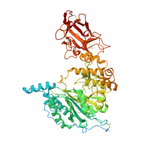
Molecular basis for fibroblast growth factor 23 O-glycosylation by GalNAc-T3.

de Las Rivas, M.Paul Daniel, E.J.Narimatsu, Y.Companon, I.Kato, K.Hermosilla, P.Thureau, A.Ceballos-Laita, L.Coelho, H.Bernado, P.Marcelo, F.Hansen, L.Maeda, R.Lostao, A.Corzana, F.Clausen, H.Gerken, T.A.Hurtado-Guerrero, R.

(2020) Nat Chem Biol 16: 351-360

  • DOI: https://doi.org/10.1038/s41589-019-0444-x
  • Primary Citation Related Structures: 
    6S22, 6S24

  • PubMed Abstract: 

    Polypeptide GalNAc-transferase T3 (GalNAc-T3) regulates fibroblast growth factor 23 (FGF23) by O-glycosylating Thr178 in a furin proprotein processing motif RHT 178 R↓S. FGF23 regulates phosphate homeostasis and deficiency in GALNT3 or FGF23 results in hyperphosphatemia and familial tumoral calcinosis. We explored the molecular mechanism for GalNAc-T3 glycosylation of FGF23 using engineered cell models and biophysical studies including kinetics, molecular dynamics and X-ray crystallography of GalNAc-T3 complexed to glycopeptide substrates. GalNAc-T3 uses a lectin domain mediated mechanism to glycosylate Thr178 requiring previous glycosylation at Thr171. Notably, Thr178 is a poor substrate site with limiting glycosylation due to substrate clashes leading to destabilization of the catalytic domain flexible loop. We suggest GalNAc-T3 specificity for FGF23 and its ability to control circulating levels of intact FGF23 is achieved by FGF23 being a poor substrate. GalNAc-T3's structure further reveals the molecular bases for reported disease-causing mutations. Our findings provide an insight into how GalNAc-T isoenzymes achieve isoenzyme-specific nonredundant functions.


  • Organizational Affiliation
    • BIFI, University of Zaragoza, Mariano Esquillor s/n, Campus Rio Ebro, Edificio I+D, Zaragoza, Spain.

Macromolecule Content 

  • Total Structure Weight: 76.04 kDa 
  • Atom Count: 4,756 
  • Modeled Residue Count: 551 
  • Deposited Residue Count: 645 
  • Unique protein chains: 2

Macromolecules

Find similar proteins by:|  3D Structure
Entity ID: 1
MoleculeChains  Sequence LengthOrganismDetailsImage
Polypeptide N-acetylgalactosaminyltransferase631Taeniopygia guttataMutation(s): 0 
Gene Names: GALNT3
EC: 2.4.1 (PDB Primary Data), 2.4.1.41 (UniProt)
UniProt
Find proteins for H0ZAB5 (Taeniopygia guttata)
Explore H0ZAB5 
Go to UniProtKB:  H0ZAB5
Entity Groups
Sequence Clusters30% Identity50% Identity70% Identity90% Identity95% Identity100% Identity
UniProt GroupH0ZAB5
Glycosylation
Glycosylation Sites: 1
Sequence Annotations
Expand
Reference Sequence
Find similar proteins by:  Sequence   |   3D Structure  
Entity ID: 2
MoleculeChains  Sequence LengthOrganismDetailsImage
ALA-THR-GLY-ALA-GLY-ALA-GLY-ALA-GLY-THR-THR-PRO-GLY-PROB [auth F]14Homo sapiensMutation(s): 0 

Small Molecules

Ligands 6 Unique
IDChains Name / Formula / InChI Key2D Diagram3D Interactions
UDP

Query on UDP


Download:Ideal Coordinates CCD File
S [auth A]URIDINE-5'-DIPHOSPHATE
C9 H14 N2 O12 P2
XCCTYIAWTASOJW-XVFCMESISA-N
NGA
(Subject of Investigation/LOI)

Query on NGA


Download:Ideal Coordinates CCD File
V [auth F]2-acetamido-2-deoxy-beta-D-galactopyranose
C8 H15 N O6
OVRNDRQMDRJTHS-JAJWTYFOSA-N
NAG

Query on NAG


Download:Ideal Coordinates CCD File
U [auth A]2-acetamido-2-deoxy-beta-D-glucopyranose
C8 H15 N O6
OVRNDRQMDRJTHS-FMDGEEDCSA-N
TAM

Query on TAM


Download:Ideal Coordinates CCD File
C [auth A]TRIS(HYDROXYETHYL)AMINOMETHANE
C7 H17 N O3
GKODZWOPPOTFGA-UHFFFAOYSA-N
PO4

Query on PO4


Download:Ideal Coordinates CCD File
D [auth A]
E [auth A]
F [auth A]
G [auth A]
H [auth A]
D [auth A],
E [auth A],
F [auth A],
G [auth A],
H [auth A],
I [auth A],
J [auth A],
K [auth A],
L [auth A],
M [auth A],
N [auth A],
O [auth A],
P [auth A],
Q [auth A],
R [auth A],
W [auth F]
PHOSPHATE ION
O4 P
NBIIXXVUZAFLBC-UHFFFAOYSA-K
MN

Query on MN


Download:Ideal Coordinates CCD File
T [auth A]MANGANESE (II) ION
Mn
WAEMQWOKJMHJLA-UHFFFAOYSA-N

Experimental Data & Validation

Experimental Data

  • Method: X-RAY DIFFRACTION
  • Resolution: 2.12 Å
  • R-Value Free:  0.237 (Depositor), 0.237 (DCC) 
  • R-Value Work:  0.191 (Depositor), 0.199 (DCC) 
  • R-Value Observed: 0.193 (Depositor) 
Space Group: P 21 21 21
Unit Cell:
Length ( Å )Angle ( ˚ )
a = 50.655α = 90
b = 104.661β = 90
c = 143.186γ = 90
Software Package:
Software NamePurpose
REFMACrefinement
XDSdata reduction
SCALAdata scaling
PHASERphasing

Structure Validation

View Full Validation Report


Ligand Structure Quality Assessment 


Entry History 

Revision History  (Full details and data files)

  • Version 1.0: 2019-12-25
    Type: Initial release
  • Version 1.1: 2020-01-22
    Changes: Database references
  • Version 1.2: 2020-03-04
    Changes: Data collection, Database references
  • Version 1.3: 2020-07-29
    Type: Remediation
    Reason: Carbohydrate remediation
    Changes: Data collection, Derived calculations, Structure summary
  • Version 1.4: 2024-01-24
    Changes: Data collection, Database references, Refinement description, Structure summary
  • Version 1.5: 2024-11-06
    Changes: Structure summary