6S0H | pdb_00006s0h

Structure of IMP-13 metallo-beta-lactamase complexed with hydrolysed doripenem


Experimental Data Snapshot

  • Method: X-RAY DIFFRACTION
  • Resolution: 2.85 Å
  • R-Value Free: 
    0.263 (Depositor), 0.265 (DCC) 
  • R-Value Work: 
    0.228 (Depositor), 0.228 (DCC) 
  • R-Value Observed: 
    0.230 (Depositor) 

wwPDB Validation 3D Report Full Report

Validation slider image for 6S0H

Ligand Structure Quality Assessment 


This is version 1.4 of the entry. See complete history

Literature

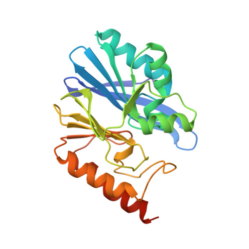
Structure and Molecular Recognition Mechanism of IMP-13 Metallo-beta-Lactamase.

Softley, C.A.Zak, K.M.Bostock, M.J.Fino, R.Zhou, R.X.Kolonko, M.Mejdi-Nitiu, R.Meyer, H.Sattler, M.Popowicz, G.M.

(2020) Antimicrob Agents Chemother 64

  • DOI: https://doi.org/10.1128/AAC.00123-20
  • Primary Citation Related Structures: 
    6R73, 6R78, 6R79, 6RZR, 6RZS, 6S0H

  • PubMed Abstract: 

    Multidrug resistance among Gram-negative bacteria is a major global public health threat. Metallo-β-lactamases (MBLs) target the most widely used antibiotic class, the β-lactams, including the most recent generation of carbapenems. Interspecies spread renders these enzymes a serious clinical threat, and there are no clinically available inhibitors. We present the crystal structures of IMP-13, a structurally uncharacterized MBL from the Gram-negative bacterium Pseudomonas aeruginosa found in clinical outbreaks globally, and characterize the binding using solution nuclear magnetic resonance spectroscopy and molecular dynamics simulations. The crystal structures of apo IMP-13 and IMP-13 bound to four clinically relevant carbapenem antibiotics (doripenem, ertapenem, imipenem, and meropenem) are presented. Active-site plasticity and the active-site loop, where a tryptophan residue stabilizes the antibiotic core scaffold, are essential to the substrate-binding mechanism. The conserved carbapenem scaffold plays the most significant role in IMP-13 binding, explaining the broad substrate specificity. The observed plasticity and substrate-locking mechanism provide opportunities for rational drug design of novel metallo-β-lactamase inhibitors, essential in the fight against antibiotic resistance.


  • Organizational Affiliation
    • Biomolecular NMR and Center for Integrated Protein Science Munich at Department Chemie, Technical University of Munich, Garching, Germany.

Macromolecule Content 

  • Total Structure Weight: 51.97 kDa 
  • Atom Count: 3,541 
  • Modeled Residue Count: 432 
  • Deposited Residue Count: 454 
  • Unique protein chains: 1

Macromolecules

Find similar proteins by:|  3D Structure
Entity ID: 1
MoleculeChains  Sequence LengthOrganismDetailsImage
Beta-lactamase
A, B
227Pseudomonas aeruginosaMutation(s): 0 
Gene Names: bla-imp13bla-IMP13blaIMP-13
EC: 3.5.2.6
UniProt
Find proteins for Q7WYA8 (Pseudomonas aeruginosa)
Explore Q7WYA8 
Go to UniProtKB:  Q7WYA8
Entity Groups
Sequence Clusters30% Identity50% Identity70% Identity90% Identity95% Identity100% Identity
UniProt GroupQ7WYA8
Sequence Annotations
Expand
Reference Sequence

Small Molecules

Ligands 3 Unique
IDChains Name / Formula / InChI Key2D Diagram3D Interactions
KQ8
(Subject of Investigation/LOI)

Query on KQ8


Download:Ideal Coordinates CCD File
L [auth A],
P [auth B]
(2~{R},3~{R})-2-[(2~{S},3~{R})-1,3-bis(oxidanyl)-1-oxidanylidene-butan-2-yl]-3-methyl-4-[(3~{S},5~{S})-5-[(sulfamoylamino)methyl]pyrrolidin-3-yl]sulfanyl-2,3-dihydro-1~{H}-pyrrole-5-carboxylic acid
C15 H26 N4 O7 S2
DGCDKVKIMXIBKA-ZBJKAAEASA-N
ZN

Query on ZN


Download:Ideal Coordinates CCD File
C [auth A],
D [auth A],
N [auth B],
O [auth B]
ZINC ION
Zn
PTFCDOFLOPIGGS-UHFFFAOYSA-N
EDO

Query on EDO


Download:Ideal Coordinates CCD File
E [auth A]
F [auth A]
G [auth A]
H [auth A]
I [auth A]
E [auth A],
F [auth A],
G [auth A],
H [auth A],
I [auth A],
J [auth A],
K [auth A],
M [auth B]
1,2-ETHANEDIOL
C2 H6 O2
LYCAIKOWRPUZTN-UHFFFAOYSA-N

Experimental Data & Validation

Experimental Data

  • Method: X-RAY DIFFRACTION
  • Resolution: 2.85 Å
  • R-Value Free:  0.263 (Depositor), 0.265 (DCC) 
  • R-Value Work:  0.228 (Depositor), 0.228 (DCC) 
  • R-Value Observed: 0.230 (Depositor) 
Space Group: P 1 21 1
Unit Cell:
Length ( Å )Angle ( ˚ )
a = 61.43α = 90
b = 72.318β = 111.72
c = 62.933γ = 90
Software Package:
Software NamePurpose
REFMACrefinement
XDSdata reduction
Aimlessdata scaling
PHASERphasing
PDB_EXTRACTdata extraction

Structure Validation

View Full Validation Report


Ligand Structure Quality Assessment 


Entry History 

& Funding Information

Deposition Data


Funding OrganizationLocationGrant Number
European UnionGermany675555
German Federal Ministry for Education and ResearchGermanyGFTARV38
Polish National Science CentrePoland2018/28/T/NZ1/00337

Revision History  (Full details and data files)

  • Version 1.0: 2020-04-01
    Type: Initial release
  • Version 1.1: 2020-04-15
    Changes: Structure summary
  • Version 1.2: 2020-04-29
    Changes: Database references
  • Version 1.3: 2020-06-03
    Changes: Database references
  • Version 1.4: 2024-05-15
    Changes: Data collection, Database references, Derived calculations