6RYM | pdb_00006rym

Structure of carbohydrate recognition domain with GlcNAc bound


Experimental Data Snapshot

  • Method: X-RAY DIFFRACTION
  • Resolution: 1.46 Å
  • R-Value Free: 
    0.184 (Depositor), 0.190 (DCC) 
  • R-Value Work: 
    0.161 (Depositor), 0.170 (DCC) 
  • R-Value Observed: 
    0.163 (Depositor) 

Starting Model: experimental
View more details

wwPDB Validation   3D Report Full Report


Ligand Structure Quality Assessment 

Created with Raphaël 2.3.0Worse 01 BetterLigand structure goodness of fit to experimental dataBest fitted NAGClick on this verticalbar to view detailsBest fitted NDGClick on this verticalbar to view details

This is version 2.2 of the entry. See complete history


Literature

Atomic-resolution crystal structures of the immune protein conglutinin from cow reveal specific interactions of its binding site withN-acetylglucosamine.

Paterson, J.M.Shaw, A.J.Burns, I.Dodds, A.W.Prasad, A.Reid, K.B.Greenhough, T.J.Shrive, A.K.

(2019) J Biological Chem 294: 17155-17165

  • DOI: https://doi.org/10.1074/jbc.RA119.010271
  • Primary Citation of Related Structures:  
    6RYG, 6RYJ, 6RYM, 6RYN

  • PubMed Abstract: 

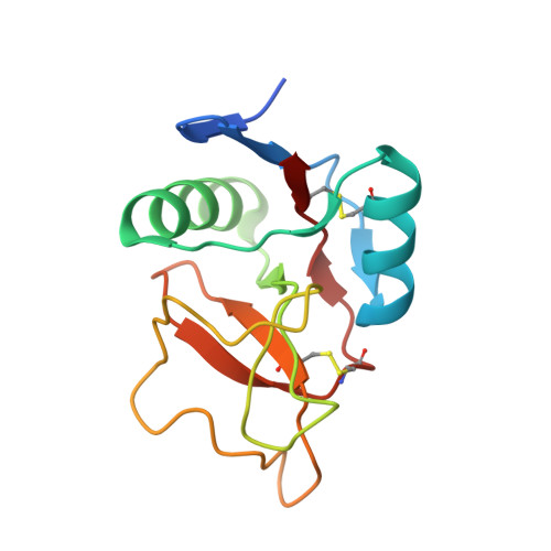
    Bovine conglutinin is an immune protein that is involved in host resistance to microbes and parasites and interacts with complement component iC3b, agglutinates erythrocytes, and neutralizes influenza A virus. Here, we determined the high-resolution (0.97-1.46 Å) crystal structures with and without bound ligand of a recombinant fragment of conglutinin's C-terminal carbohydrate-recognition domain (CRD). The structures disclosed that the high-affinity ligand N -acetyl-d-glucosamine (GlcNAc) binds in the collectin CRD calcium site by interacting with the O3' and O4' hydroxyls alongside additional specific interactions of the N -acetyl group oxygen and nitrogen with Lys-343 and Asp-320, respectively. These residues, unique to conglutinin and differing both in sequence and in location from those in other collectins, result in specific, high-affinity binding for GlcNAc. The binding pocket flanking residue Val-339, unlike the equivalent Arg-343 in the homologous human surfactant protein D, is sufficiently small to allow conglutinin Lys-343 access to the bound ligand, whereas Asp-320 lies in an extended loop proximal to the ligand-binding site and bounded at both ends by conserved residues that coordinate to both calcium and ligand. This loop becomes ordered on ligand binding. The electron density revealed both α and β anomers of GlcNAc, consistent with the added α/βGlcNAc mixture. Crystals soaked with α1-2 mannobiose, a putative component of iC3b, reported to bind to conglutinin, failed to reveal bound ligand, suggesting a requirement for presentation of mannobiose as part of an extended physiological ligand. These results reveal a highly specific GlcNAc-binding pocket in conglutinin and a novel collectin mode of carbohydrate recognition.


  • Organizational Affiliation

    School of Life Sciences, Keele University, Staffordshire ST5 5BG, United Kingdom.


Macromolecules
Find similar proteins by:  (by identity cutoff)  |  3D Structure
Entity ID: 1
MoleculeChains Sequence LengthOrganismDetailsImage
Conglutinin128Bos taurusMutation(s): 0 
Gene Names: CGN1
UniProt
Find proteins for P23805 (Bos taurus)
Explore P23805 
Go to UniProtKB:  P23805
Entity Groups  
Sequence Clusters30% Identity50% Identity70% Identity90% Identity95% Identity100% Identity
UniProt GroupP23805
Sequence Annotations
Expand
  • Reference Sequence
Experimental Data & Validation

Experimental Data

  • Method: X-RAY DIFFRACTION
  • Resolution: 1.46 Å
  • R-Value Free:  0.184 (Depositor), 0.190 (DCC) 
  • R-Value Work:  0.161 (Depositor), 0.170 (DCC) 
  • R-Value Observed: 0.163 (Depositor) 
Space Group: P 43
Unit Cell:
Length ( Å )Angle ( ˚ )
a = 50.225α = 90
b = 50.225β = 90
c = 51.925γ = 90
Software Package:
Software NamePurpose
MOSFLMdata reduction
SCALAdata scaling
REFMACrefinement
PDB_EXTRACTdata extraction
REFMACphasing

Structure Validation

View Full Validation Report


Ligand Structure Quality Assessment 

Created with Raphaël 2.3.0Worse 01 BetterLigand structure goodness of fit to experimental dataBest fitted NAGClick on this verticalbar to view detailsBest fitted NDGClick on this verticalbar to view details

Entry History 

Deposition Data

Revision History  (Full details and data files)

  • Version 1.0: 2019-10-09
    Type: Initial release
  • Version 1.1: 2019-11-20
    Changes: Database references
  • Version 2.0: 2020-07-29
    Type: Remediation
    Reason: Carbohydrate remediation
    Changes: Atomic model, Data collection, Derived calculations, Structure summary
  • Version 2.1: 2024-01-24
    Changes: Data collection, Database references, Refinement description, Structure summary
  • Version 2.2: 2024-11-06
    Changes: Structure summary