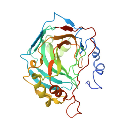
6RW1

Crystal structure of hCA II in complex with Urea, N-(1,3-dihydro-1-hydroxy-2,1-benzoxaborol-6-yl)-N'-(phenylmethyl)-


Experimental Data Snapshot

  • Method: X-RAY DIFFRACTION
  • Resolution: 1.70 Å
  • R-Value Free: 0.205 
  • R-Value Work: 0.179 

Starting Model: experimental
View more details

wwPDB Validation   3D Report Full Report


Ligand Structure Quality Assessment 


This is version 2.1 of the entry. See complete history


Literature

Exploring benzoxaborole derivatives as carbonic anhydrase inhibitors: a structural and computational analysis reveals their conformational variability as a tool to increase enzyme selectivity.

Langella, E.Alterio, V.D'Ambrosio, K.Cadoni, R.Winum, J.Y.Supuran, C.T.Monti, S.M.De Simone, G.Di Fiore, A.

(2019) J Enzyme Inhib Med Chem 34: 1498-1505

  • DOI: https://doi.org/10.1080/14756366.2019.1653291
  • Primary Citation of Related Structures:  
    6RVF, 6RVK, 6RVL, 6RW1

  • PubMed Abstract: 

    Recent studies identified the benzoxaborole moiety as a new zinc-binding group able to interact with carbonic anhydrase (CA) active site. Here, we report a structural analysis of benzoxaboroles containing urea/thiourea groups, showing that these molecules are very versatile since they can bind the enzyme assuming different binding conformations and coordination geometries of the catalytic zinc ion. In addition, theoretical calculations of binding free energy were performed highlighting the key role of specific residues for protein-inhibitor recognition. Overall, these data are very useful for the development of new inhibitors with higher selectivity and efficacy for various CAs.


  • Organizational Affiliation

    Istituto di Biostrutture e Bioimmagini, Consiglio Nazionale delle Ricerche , Naples , Italy.


Macromolecules
Find similar proteins by:  (by identity cutoff)  |  3D Structure
Entity ID: 1
MoleculeChains Sequence LengthOrganismDetailsImage
Carbonic anhydrase 2262Homo sapiensMutation(s): 0 
Gene Names: CA2
EC: 4.2.1.1 (PDB Primary Data), 4.2.1.69 (UniProt)
UniProt & NIH Common Fund Data Resources
Find proteins for P00918 (Homo sapiens)
Explore P00918 
Go to UniProtKB:  P00918
PHAROS:  P00918
GTEx:  ENSG00000104267 
Entity Groups  
Sequence Clusters30% Identity50% Identity70% Identity90% Identity95% Identity100% Identity
UniProt GroupP00918
Sequence Annotations
Expand
  • Reference Sequence
Small Molecules
Ligands 2 Unique
IDChains Name / Formula / InChI Key2D Diagram3D Interactions
KL5 (Subject of Investigation/LOI)
Query on KL5

Download Ideal Coordinates CCD File 
C [auth A]1-[7,7-bis(oxidanyl)-8-oxa-7-boranuidabicyclo[4.3.0]nona-1(6),2,4-trien-4-yl]-3-(phenylmethyl)thiourea
C15 H16 B N2 O3 S
PURMMQOEOOZDEQ-UHFFFAOYSA-N
ZN
Query on ZN

Download Ideal Coordinates CCD File 
B [auth A]ZINC ION
Zn
PTFCDOFLOPIGGS-UHFFFAOYSA-N
Experimental Data & Validation

Experimental Data

  • Method: X-RAY DIFFRACTION
  • Resolution: 1.70 Å
  • R-Value Free: 0.205 
  • R-Value Work: 0.179 
  • Space Group: P 1 21 1
Unit Cell:
Length ( Å )Angle ( ˚ )
a = 42.42α = 90
b = 41.49β = 104.54
c = 72.13γ = 90
Software Package:
Software NamePurpose
CNSrefinement
SCALEPACKdata scaling
PDB_EXTRACTdata extraction
DENZOdata reduction
CNSphasing

Structure Validation

View Full Validation Report


Ligand Structure Quality Assessment 


Entry History 

Deposition Data

Revision History  (Full details and data files)

  • Version 1.0: 2019-08-28
    Type: Initial release
  • Version 2.0: 2019-10-30
    Changes: Data collection, Derived calculations, Non-polymer description, Structure summary
  • Version 2.1: 2024-01-24
    Changes: Data collection, Database references, Refinement description