6RTE

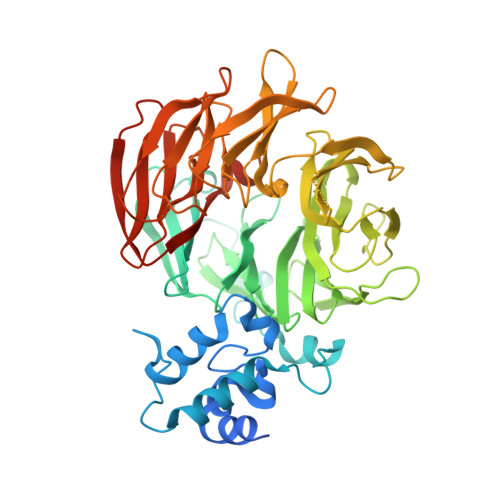
Dihydro-heme d1 dehydrogenase NirN in complex with DHE


Experimental Data Snapshot

  • Method: X-RAY DIFFRACTION
  • Resolution: 1.94 Å
  • R-Value Free: 0.198 
  • R-Value Work: 0.167 
  • R-Value Observed: 0.169 

Starting Model: experimental
View more details

wwPDB Validation   3D Report Full Report


Ligand Structure Quality Assessment 


This is version 1.4 of the entry. See complete history


Literature

Crystal Structure of Dihydro-Heme d1Dehydrogenase NirN from Pseudomonas aeruginosa Reveals Amino Acid Residues Essential for Catalysis.

Klunemann, T.Preuss, A.Adamczack, J.Rosa, L.F.M.Harnisch, F.Layer, G.Blankenfeldt, W.

(2019) J Mol Biol 431: 3246-3260

  • DOI: https://doi.org/10.1016/j.jmb.2019.05.046
  • Primary Citation of Related Structures:  
    6RTD, 6RTE

  • PubMed Abstract: 

    Many bacteria can switch from oxygen to nitrogen oxides, such as nitrate or nitrite, as terminal electron acceptors in their respiratory chain. This process is called "denitrification" and enables biofilm formation of the opportunistic human pathogen Pseudomonas aeruginosa, making it more resilient to antibiotics and highly adaptable to different habitats. The reduction of nitrite to nitric oxide is a crucial step during denitrification. It is catalyzed by the homodimeric cytochrome cd 1 nitrite reductase (NirS), which utilizes the unique isobacteriochlorin heme d 1 as its reaction center. Although the reaction mechanism of nitrite reduction is well understood, far less is known about the biosynthesis of heme d 1 . The last step of its biosynthesis introduces a double bond in a propionate group of the tetrapyrrole to form an acrylate group. This conversion is catalyzed by the dehydrogenase NirN via a unique reaction mechanism. To get a more detailed insight into this reaction, the crystal structures of NirN with and without bound substrate have been determined. Similar to the homodimeric NirS, the monomeric NirN consists of an eight-bladed heme d 1 -binding β-propeller and a cytochrome c domain, but their relative orientation differs with respect to NirS. His147 coordinates heme d 1 at the proximal side, whereas His323, which belongs to a flexible loop, binds at the distal position. Tyr461 and His417 are located next to the hydrogen atoms removed during dehydrogenation, suggesting an important role in catalysis. Activity assays with NirN variants revealed the essentiality of His147, His323 and Tyr461, but not of His417.


  • Organizational Affiliation

    Department of Structure and Function of Proteins, Helmholtz Centre for Infection Research, Inhoffenstrasse 7, 38124 Braunschweig, Germany.


Macromolecules
Find similar proteins by:  (by identity cutoff)  |  3D Structure
Entity ID: 1
MoleculeChains Sequence LengthOrganismDetailsImage
Cytochrome c
A, B
509Pseudomonas aeruginosaMutation(s): 0 
Gene Names: nirS_1EFK27_00335EGV95_02815IPC669_26980PAERUG_E15_London_28_01_14_07983RW109_RW109_01113
EC: 1.7.2.1 (PDB Primary Data), 1.3 (UniProt)
UniProt
Find proteins for Q9I609 (Pseudomonas aeruginosa (strain ATCC 15692 / DSM 22644 / CIP 104116 / JCM 14847 / LMG 12228 / 1C / PRS 101 / PAO1))
Explore Q9I609 
Go to UniProtKB:  Q9I609
Entity Groups  
Sequence Clusters30% Identity50% Identity70% Identity90% Identity95% Identity100% Identity
UniProt GroupQ9I609
Sequence Annotations
Expand
  • Reference Sequence
Experimental Data & Validation

Experimental Data

  • Method: X-RAY DIFFRACTION
  • Resolution: 1.94 Å
  • R-Value Free: 0.198 
  • R-Value Work: 0.167 
  • R-Value Observed: 0.169 
  • Space Group: C 1 2 1
Unit Cell:
Length ( Å )Angle ( ˚ )
a = 191.123α = 90
b = 54.957β = 132.417
c = 131.896γ = 90
Software Package:
Software NamePurpose
autoPROCdata collection
PHENIXrefinement
XDSdata reduction
Aimlessdata scaling
Cootmodel building
BALBESphasing

Structure Validation

View Full Validation Report


Ligand Structure Quality Assessment 


Entry History & Funding Information

Deposition Data


Funding OrganizationLocationGrant Number
German Research FoundationGermanyGRK 2223
German Research FoundationGermanyLA 2412

Revision History  (Full details and data files)

  • Version 1.0: 2019-06-19
    Type: Initial release
  • Version 1.1: 2019-08-28
    Changes: Data collection, Database references
  • Version 1.2: 2019-11-20
    Changes: Derived calculations
  • Version 1.3: 2024-01-24
    Changes: Data collection, Database references, Derived calculations, Refinement description
  • Version 1.4: 2024-10-23
    Changes: Structure summary