


Endothiapepsin in complex with 017
Magari, F., Heine, A., Konstantinidou, M., Sutanto, F., Haupenthal, J., Jumde, R.V., Unver, M.Y., Camacho, C.J., Hirsch, A.K.H., Doemling, A., Klebe, G.To be published.
Experimental Data Snapshot
Entity ID: 1 | |||||
|---|---|---|---|---|---|
| Molecule | Chains | Sequence Length | Organism | Details | Image |
| Endothiapepsin | 330 | Cryphonectria parasitica | Mutation(s): 0 EC: 3.4.23.22 | 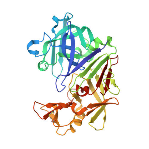 | |
UniProt | |||||
Entity Groups | |||||
| Sequence Clusters | 30% Identity50% Identity70% Identity90% Identity95% Identity100% Identity | ||||
| UniProt Group | P11838 | ||||
Sequence AnnotationsExpand | |||||
Reference Sequence | |||||
| Ligands 4 Unique | |||||
|---|---|---|---|---|---|
| ID | Chains | Name / Formula / InChI Key | 2D Diagram | 3D Interactions | |
| KHW Download:Ideal Coordinates CCD File | C [auth A] | [(1~{R})-1-[1-(phenylmethyl)-1,2,3,4-tetrazol-5-yl]butyl]diazane C12 H18 N6 RXFMIKHNJVMPGC-LLVKDONJSA-N |  | ||
| KHZ Download:Ideal Coordinates CCD File | E [auth A] | 1~{H}-1,2,3,4-tetrazol-5-ylmethyldiazane C2 H6 N6 LKTRPEVOGNHGTR-UHFFFAOYSA-N |  | ||
| GOL Download:Ideal Coordinates CCD File | B [auth A] | GLYCEROL C3 H8 O3 PEDCQBHIVMGVHV-UHFFFAOYSA-N |  | ||
| DMS Download:Ideal Coordinates CCD File | D [auth A] | DIMETHYL SULFOXIDE C2 H6 O S IAZDPXIOMUYVGZ-UHFFFAOYSA-N |  | ||
| Length ( Å ) | Angle ( ˚ ) |
|---|---|
| a = 45.4 | α = 90 |
| b = 73.269 | β = 109.75 |
| c = 52.947 | γ = 90 |
| Software Name | Purpose |
|---|---|
| PHENIX | refinement |
| MxCuBE | data collection |
| XDS | data scaling |
| XDS | data reduction |
| PHASER | phasing |


