6RSK | pdb_00006rsk

Cytochrome c co-crystallized with 20 eq. sulfonato-calix[8]arene and 15 eq. spermine - structure II


Experimental Data Snapshot

  • Method: X-RAY DIFFRACTION
  • Resolution: 2.31 Å
  • R-Value Free: 
    0.222 (Depositor), 0.252 (DCC) 
  • R-Value Work: 
    0.183 (Depositor) 
  • R-Value Observed: 
    0.185 (Depositor) 

Starting Model: experimental
View more details

wwPDB Validation 3D Report Full Report

Validation slider image for 6RSK

Ligand Structure Quality Assessment 


This is version 1.3 of the entry. See complete history

Literature

Tuning Protein Frameworks via Auxiliary Supramolecular Interactions.

Engilberge, S.Rennie, M.L.Dumont, E.Crowley, P.B.

(2019) ACS Nano 13: 10343-10350

  • DOI: https://doi.org/10.1021/acsnano.9b04115
  • Primary Citation Related Structures: 
    6GDA, 6RSI, 6RSJ, 6RSK, 6RSL

  • PubMed Abstract: 

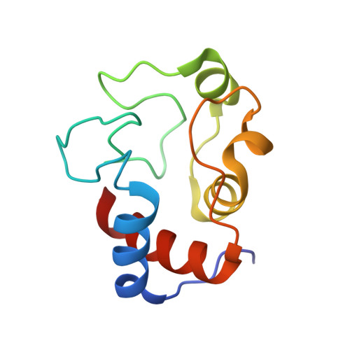
    Protein crystals with their precise, periodic array of functional building blocks have potential applications in biomaterials, sensing, and catalysis. This paper describes how a highly porous crystalline framework of a cationic redox protein and an anionic macrocycle can be modulated by a small cationic effector. Ternary composites of protein (∼13 kDa), calix[8]arene (∼1.5 kDa), and effector (∼0.2 kDa) formed distinct crystalline architectures, dependent on the effector concentration and the crystallization technique. A combination of X-ray crystallography and density functional theory (DFT) calculations was used to decipher the framework variations, which appear to be dependent on a calixarene conformation change mediated by the effector. This "switch" calixarene was observed in three states, each of which is associated with a different interaction network. Two structures obtained by co-crystallization with the effector contained an additional protein "pillar", resulting in framework duplication and decreased porosity. These results suggest how protein assembly can be engineered by supramolecular host-guest interactions.


  • Organizational Affiliation
    • School of Chemistry , National University of Ireland Galway , University Road , Galway H91 TK33 , Ireland.

Macromolecule Content 

  • Total Structure Weight: 36.12 kDa 
  • Atom Count: 2,644 
  • Modeled Residue Count: 216 
  • Deposited Residue Count: 216 
  • Unique protein chains: 1

Macromolecules

Find similar proteins by:|  3D Structure
Entity ID: 1
MoleculeChains  Sequence LengthOrganismDetailsImage
Cytochrome c iso-1
A, B
108Saccharomyces cerevisiae S288CMutation(s): 1 
Gene Names: CYC1YJR048WJ1653
UniProt
Find proteins for P00044 (Saccharomyces cerevisiae (strain ATCC 204508 / S288c))
Explore P00044 
Go to UniProtKB:  P00044
Entity Groups
Sequence Clusters30% Identity50% Identity70% Identity90% Identity95% Identity100% Identity
UniProt GroupP00044
Sequence Annotations
Expand
Reference Sequence

Small Molecules

Ligands 4 Unique
IDChains Name / Formula / InChI Key2D Diagram3D Interactions
EVB
(Subject of Investigation/LOI)

Query on EVB


Download:Ideal Coordinates CCD File
D [auth A]
E [auth A]
F [auth A]
P [auth B]
Q [auth B]
D [auth A],
E [auth A],
F [auth A],
P [auth B],
Q [auth B],
R [auth B]
sulfonato-calix[8]arene
C56 H48 O32 S8
KCEGJGDGMRAJEP-UHFFFAOYSA-N
HEC

Query on HEC


Download:Ideal Coordinates CCD File
C [auth A],
O [auth B]
HEME C
C34 H34 Fe N4 O4
HXQIYSLZKNYNMH-LJNAALQVSA-N
SPM
(Subject of Investigation/LOI)

Query on SPM


Download:Ideal Coordinates CCD File
M [auth A],
N [auth A],
X [auth B],
Y [auth B]
SPERMINE
C10 H26 N4
PFNFFQXMRSDOHW-UHFFFAOYSA-N
SO4

Query on SO4


Download:Ideal Coordinates CCD File
G [auth A]
H [auth A]
I [auth A]
J [auth A]
K [auth A]
G [auth A],
H [auth A],
I [auth A],
J [auth A],
K [auth A],
L [auth A],
S [auth B],
T [auth B],
U [auth B],
V [auth B],
W [auth B]
SULFATE ION
O4 S
QAOWNCQODCNURD-UHFFFAOYSA-L

Experimental Data & Validation

Experimental Data

  • Method: X-RAY DIFFRACTION
  • Resolution: 2.31 Å
  • R-Value Free:  0.222 (Depositor), 0.252 (DCC) 
  • R-Value Work:  0.183 (Depositor) 
  • R-Value Observed: 0.185 (Depositor) 
Space Group: P 43 21 2
Unit Cell:
Length ( Å )Angle ( ˚ )
a = 100.459α = 90
b = 100.459β = 90
c = 89.539γ = 90
Software Package:
Software NamePurpose
BUSTERrefinement
XDSdata reduction
Aimlessdata scaling
PHASERphasing

Structure Validation

View Full Validation Report


Ligand Structure Quality Assessment 


Entry History 

& Funding Information

Deposition Data


Funding OrganizationLocationGrant Number
Science Foundation IrelandIreland13/CDA/2168

Revision History  (Full details and data files)

  • Version 1.0: 2019-09-18
    Type: Initial release
  • Version 1.1: 2019-10-23
    Changes: Data collection, Database references
  • Version 1.2: 2024-01-24
    Changes: Data collection, Database references, Derived calculations, Refinement description
  • Version 1.3: 2025-10-01
    Changes: Advisory, Derived calculations, Structure summary