6ROH | pdb_00006roh

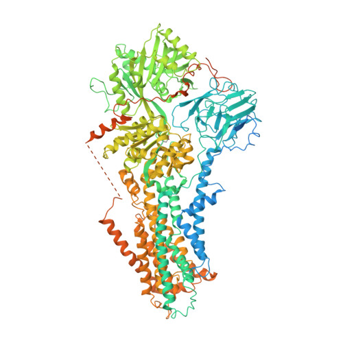
Cryo-EM structure of the autoinhibited Drs2p-Cdc50p


Experimental Data Snapshot

  • Method: ELECTRON MICROSCOPY
  • Resolution: 2.80 Å
  • Aggregation State: PARTICLE 
  • Reconstruction Method: SINGLE PARTICLE 

wwPDB Validation 3D Report Full Report

Validation slider image for 6ROH

This is version 3.0 of the entry. See complete history. 

Literature

Structure and autoregulation of a P4-ATPase lipid flippase.

Timcenko, M., Lyons, J.A., Januliene, D., Ulstrup, J.J., Dieudonne, T., Montigny, C., Ash, M.R., Karlsen, J.L., Boesen, T., Kuhlbrandt, W., Lenoir, G., Moeller, A., Nissen, P.

(2019) Nature 571: 366-370

  • DOI: https://doi.org/10.1038/s41586-019-1344-7
  • Primary Citation Related Structures: 
    6ROH, 6ROI, 6ROJ

  • PubMed Abstract: 

    Type 4 P-type ATPases (P4-ATPases) are lipid flippases that drive the active transport of phospholipids from exoplasmic or luminal leaflets to cytosolic leaflets of eukaryotic membranes. The molecular architecture of P4-ATPases and the mechanism through which they recognize and transport lipids have remained unknown. Here we describe the cryo-electron microscopy structure of the P4-ATPase Drs2p-Cdc50p, a Saccharomyces cerevisiae lipid flippase that is specific to phosphatidylserine and phosphatidylethanolamine. Drs2p-Cdc50p is autoinhibited by the C-terminal tail of Drs2p, and activated by the lipid phosphatidylinositol-4-phosphate (PtdIns4P or PI4P). We present three structures that represent the complex in an autoinhibited, an intermediate and a fully activated state. The analysis highlights specific features of P4-ATPases and reveals sites of autoinhibition and PI4P-dependent activation. We also observe a putative lipid translocation pathway in this flippase that involves a conserved PISL motif in transmembrane segment 4 and polar residues of transmembrane segments 2 and 5, in particular Lys1018, in the centre of the lipid bilayer.


  • Organizational Affiliation: 
    • DANDRITE, Nordic EMBL Partnership for Molecular Medicine, Department of Molecular Biology and Genetics, Aarhus University, Aarhus, Denmark.

Macromolecule Content 

  • Total Structure Weight: 212.98 kDa 
  • Atom Count: 11,758 
  • Modeled Residue Count: 1,449 
  • Deposited Residue Count: 1,873 
  • Unique protein chains: 2

Macromolecules

Find similar proteins by:|  3D Structure
Entity ID: 1
MoleculeChains  Sequence LengthOrganismDetailsImage
Probable phospholipid-transporting ATPase DRS21,460Saccharomyces cerevisiae S288CMutation(s): 0 
Gene Names: DRS2, YAL026C, FUN38
EC: 7.6.2.1
Membrane Entity: Yes 
UniProt
Find proteins for P39524 (Saccharomyces cerevisiae (strain ATCC 204508 / S288c))
Explore P39524 
Go to UniProtKB:  P39524
Entity Groups
Sequence Clusters30% Identity50% Identity70% Identity90% Identity95% Identity100% Identity
UniProt GroupP39524
Sequence Annotations
Expand
Reference Sequence
Find similar proteins by:|  3D Structure
Entity ID: 2
MoleculeChains  Sequence LengthOrganismDetailsImage
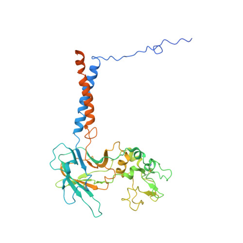
Cell division control protein 50B [auth C]413Saccharomyces cerevisiae S288CMutation(s): 0 
Gene Names: CDC50, YCR094W, YCR94W
Membrane Entity: Yes 
UniProt
Find proteins for P25656 (Saccharomyces cerevisiae (strain ATCC 204508 / S288c))
Explore P25656 
Go to UniProtKB:  P25656
Entity Groups
Sequence Clusters30% Identity50% Identity70% Identity90% Identity95% Identity100% Identity
UniProt GroupP25656
Glycosylation
Glycosylation Sites: 3Go to GlyGen: P25656-1
Sequence Annotations
Expand
Reference Sequence

Oligosaccharides

Help  
Entity ID: 3
MoleculeChains Length2D Diagram GlycosylationD Interactions
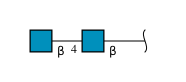
2-acetamido-2-deoxy-beta-D-glucopyranose-(1-4)-2-acetamido-2-deoxy-beta-D-glucopyranoseC [auth B]2N-Glycosylation
Glycosylation Resources
GlyTouCan: G42666HT
GlyCosmos: G42666HT
GlyGen: G42666HT
Entity ID: 4
MoleculeChains Length2D Diagram GlycosylationD Interactions
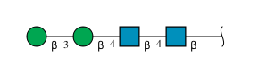
beta-D-mannopyranose-(1-3)-beta-D-mannopyranose-(1-4)-2-acetamido-2-deoxy-beta-D-glucopyranose-(1-4)-2-acetamido-2-deoxy-beta-D-glucopyranose
D
4N-Glycosylation
Glycosylation Resources
GlyTouCan: G31886NL
GlyCosmos: G31886NL
GlyGen: G31886NL

Experimental Data & Validation

Experimental Data

  • Method: ELECTRON MICROSCOPY
  • Resolution: 2.80 Å
  • Aggregation State: PARTICLE 
  • Reconstruction Method: SINGLE PARTICLE 
EM Software:
TaskSoftware PackageVersion
RECONSTRUCTIONRELION
MODEL REFINEMENTPHENIX

Structure Validation

View Full Validation Report


Entry History 

& Funding Information

Deposition Data


Funding OrganizationLocationGrant Number
Danish Council for Independent ResearchDenmark0602-02912B
LundbeckfondenDenmarkR171-2014-663
LundbeckfondenDenmarkR209-2015-2704
LundbeckfondenDenmark2015-2666
French National Research AgencyFranceANR-14-CE09-0022
French Infrastructure for Integrated Structural BiologyFranceANR-10-INSB-05

Revision History  (Full details and data files)

  • Version 1.0: 2019-07-03
    Type: Initial release
  • Version 1.1: 2019-07-10
    Changes: Data collection, Database references
  • Version 1.2: 2019-07-31
    Changes: Data collection, Database references
  • Version 1.3: 2019-12-18
    Changes: Other
  • Version 2.0: 2020-07-29
    Type: Remediation
    Reason: Carbohydrate remediation
    Changes: Atomic model, Data collection, Derived calculations, Structure summary
  • Version 2.1: 2024-10-09
    Changes: Data collection, Database references, Structure summary
  • Version 3.0: 2026-09-02
    Type: Remediation
    Reason: Metalloprotein remediation
    Changes: Atomic model, Author supporting evidence, Data collection, Derived calculations, Non-polymer description, Polymer sequence, Structure summary