6RNM | pdb_00006rnm

Crystal structure of a complex between the LlFpg protein, a THF-DNA and an inhibitor


Experimental Data Snapshot

  • Method: X-RAY DIFFRACTION
  • Resolution: 1.76 Å
  • R-Value Free: 
    0.188 (Depositor), 0.188 (DCC) 
  • R-Value Work: 
    0.169 (Depositor), 0.170 (DCC) 
  • R-Value Observed: 
    0.170 (Depositor) 

Starting Model: experimental
View more details

wwPDB Validation 3D Report Full Report

Validation slider image for 6RNM

Ligand Structure Quality Assessment 


This is version 1.1 of the entry. See complete history

Literature

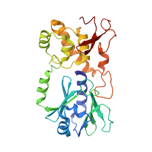
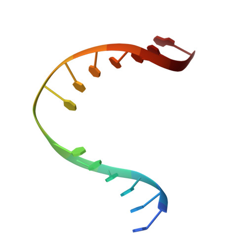
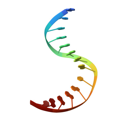
Thiopurine Derivative-Induced Fpg/Nei DNA Glycosylase Inhibition: Structural, Dynamic and Functional Insights.

Rieux, C.Goffinont, S.Coste, F.Tber, Z.Cros, J.Roy, V.Guerin, M.Gaudon, V.Bourg, S.Biela, A.Aucagne, V.Agrofoglio, L.Garnier, N.Castaing, B.

(2020) Int J Mol Sci 21

  • DOI: https://doi.org/10.3390/ijms21062058
  • Primary Citation Related Structures: 
    6RNM, 6RNO, 6RNR, 6RO2, 6ROK, 6RP0, 6RP7

  • PubMed Abstract: 

    DNA glycosylases are emerging as relevant pharmacological targets in inflammation, cancer and neurodegenerative diseases. Consequently, the search for inhibitors of these enzymes has become a very active research field. As a continuation of previous work that showed that 2-thioxanthine (2TX) is an irreversible inhibitor of zinc finger (ZnF)-containing Fpg/Nei DNA glycosylases, we designed and synthesized a mini-library of 2TX-derivatives (TXn) and evaluated their ability to inhibit Fpg/Nei enzymes. Among forty compounds, four TXn were better inhibitors than 2TX for Fpg. Unexpectedly, but very interestingly, two dithiolated derivatives more selectively and efficiently inhibit the zincless finger (ZnLF)-containing enzymes (human and mimivirus Neil1 DNA glycosylases hNeil1 and MvNei1, respectively). By combining chemistry, biochemistry, mass spectrometry, blind and flexible docking and X-ray structure analysis, we localized new TXn binding sites on Fpg/Nei enzymes. This endeavor allowed us to decipher at the atomic level the mode of action for the best TXn inhibitors on the ZnF-containing enzymes. We discovered an original inhibition mechanism for the ZnLF-containing Fpg/Nei DNA glycosylases by disulfide cyclic trimeric forms of dithiopurines. This work paves the way for the design and synthesis of a new structural class of inhibitors for selective pharmacological targeting of hNeil1 in cancer and neurodegenerative diseases.


  • Organizational Affiliation
    • Centre de Biophysique Moléculaire, UPR4301 CNRS, rue Charles Sadron, CEDEX 2, F-45071 Orléans, France.

Macromolecule Content 

  • Total Structure Weight: 39.94 kDa 
  • Atom Count: 3,240 
  • Modeled Residue Count: 299 
  • Deposited Residue Count: 299 
  • Unique protein chains: 1
  • Unique nucleic acid chains: 2

Macromolecules


Find similar proteins by:|  3D Structure
Entity ID: 1
MoleculeChains  Sequence LengthOrganismDetailsImage
Formamidopyrimidine-DNA glycosylase271Lactococcus cremorisMutation(s): 0 
Gene Names: mutMfpgNCDO763_0992
EC: 3.2.2.23 (PDB Primary Data), 4.2.99.18 (PDB Primary Data)
UniProt
Find proteins for P42371 (Lactococcus lactis subsp. cremoris)
Explore P42371 
Go to UniProtKB:  P42371
Entity Groups
Sequence Clusters30% Identity50% Identity70% Identity90% Identity95% Identity100% Identity
UniProt GroupP42371
Sequence Annotations
Expand
Reference Sequence
Find similar nucleic acids by:  Sequence
Entity ID: 2
MoleculeChains LengthOrganismImage
DNA (5'-D(*CP*TP*CP*TP*TP*TP*(3DR)P*TP*TP*TP*CP*TP*CP*G)-3')B [auth D]14synthetic construct
Sequence Annotations
Expand
Reference Sequence
Find similar nucleic acids by:  Sequence
Entity ID: 3
MoleculeChains LengthOrganismImage
DNA (5'-D(*GP*CP*GP*AP*GP*AP*AP*AP*CP*AP*AP*AP*GP*A)-3')C [auth E]14synthetic construct
Sequence Annotations
Expand
Reference Sequence

Experimental Data & Validation

Experimental Data

  • Method: X-RAY DIFFRACTION
  • Resolution: 1.76 Å
  • R-Value Free:  0.188 (Depositor), 0.188 (DCC) 
  • R-Value Work:  0.169 (Depositor), 0.170 (DCC) 
  • R-Value Observed: 0.170 (Depositor) 
Space Group: P 41 21 2
Unit Cell:
Length ( Å )Angle ( ˚ )
a = 91.957α = 90
b = 91.957β = 90
c = 142.182γ = 90
Software Package:
Software NamePurpose
XDSdata reduction
PHASERphasing
BUSTERrefinement
PDB_EXTRACTdata extraction
autoPROCdata scaling

Structure Validation

View Full Validation Report


Ligand Structure Quality Assessment 


Entry History 

Deposition Data

Revision History  (Full details and data files)

  • Version 1.0: 2020-04-15
    Type: Initial release
  • Version 1.1: 2024-01-24
    Changes: Data collection, Database references, Derived calculations, Refinement description