6RN6 | pdb_00006rn6

DPP1 in complex with inhibitor


Experimental Data Snapshot

  • Method: X-RAY DIFFRACTION
  • Resolution: 2.40 Å
  • R-Value Free: 
    0.221 (Depositor), 0.225 (DCC) 
  • R-Value Work: 
    0.174 (Depositor) 
  • R-Value Observed: 
    0.176 (Depositor) 

Starting Model: experimental
View more details

wwPDB Validation 3D Report Full Report

Validation slider image for 6RN6

Ligand Structure Quality Assessment 


This is version 1.3 of the entry. See complete history

Literature

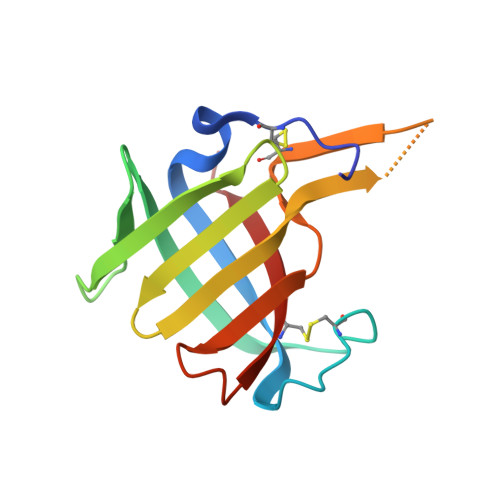
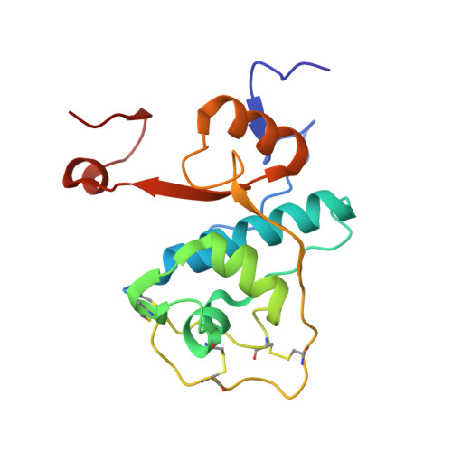
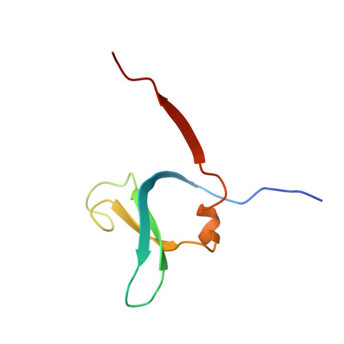
DPP1 Inhibitors: Exploring the Role of Water in the S2 Pocket of DPP1 with Substituted Pyrrolidines.

Kack, H.Doyle, K.Hughes, S.J.Bodnarchuk, M.S.Lonn, H.Van De Poel, A.Palmer, N.

(2019) ACS Med Chem Lett 10: 1222-1227

  • DOI: https://doi.org/10.1021/acsmedchemlett.9b00261
  • Primary Citation Related Structures: 
    6RN6, 6RN7, 6RN9, 6RNE, 6RNI

  • PubMed Abstract: 

    A series of pyrrolidine amino nitrile DPP1 inhibitors have been developed and characterized. The S2 pocket structure-activity relationship for these compounds shows significant gains in potency for DPP1 from interacting further with target residues and a network of water molecules in the binding pocket. Herein we describe the X-ray crystal structures of several of these compounds alongside an analysis of factors influencing the inhibitory potency toward DPP1 of which stabilization of the water network, demonstrated using Grand Canonical Monte Carlo simulations and free energy calculations, is attributed as a main factor.


  • Organizational Affiliation
    • Structure, Biophysics and Fragments, Discovery Sciences, R&D, AstraZeneca, Gothenburg, Sweden.

Macromolecule Content 

  • Total Structure Weight: 80.19 kDa 
  • Atom Count: 5,755 
  • Modeled Residue Count: 688 
  • Deposited Residue Count: 702 
  • Unique protein chains: 3

Macromolecules

Find similar proteins by:|  3D Structure
Entity ID: 1
MoleculeChains  Sequence LengthOrganismDetailsImage
Dipeptidyl peptidase 1
A, D
119Homo sapiensMutation(s): 0 
Gene Names: CTSCCPPI
EC: 3.4.14.1
UniProt & NIH Common Fund Data Resources
Find proteins for P53634 (Homo sapiens)
Explore P53634 
Go to UniProtKB:  P53634
PHAROS:  P53634
GTEx:  ENSG00000109861 
Entity Groups
Sequence Clusters30% Identity50% Identity70% Identity90% Identity95% Identity100% Identity
UniProt GroupP53634
Glycosylation
Glycosylation Sites: 2Go to GlyGen: P53634-1
Sequence Annotations
Expand
Reference Sequence
Find similar proteins by:|  3D Structure
Entity ID: 2
MoleculeChains  Sequence LengthOrganismDetailsImage
Dipeptidyl peptidase 1
B, E
163Homo sapiensMutation(s): 0 
Gene Names: CTSCCPPI
EC: 3.4.14.1
UniProt & NIH Common Fund Data Resources
Find proteins for P53634 (Homo sapiens)
Explore P53634 
Go to UniProtKB:  P53634
PHAROS:  P53634
GTEx:  ENSG00000109861 
Entity Groups
Sequence Clusters30% Identity50% Identity70% Identity90% Identity95% Identity100% Identity
UniProt GroupP53634
Glycosylation
Glycosylation Sites: 1Go to GlyGen: P53634-1
Sequence Annotations
Expand
Reference Sequence
Find similar proteins by:|  3D Structure
Entity ID: 3
MoleculeChains  Sequence LengthOrganismDetailsImage
Dipeptidyl peptidase 1
C, F
69Homo sapiensMutation(s): 0 
Gene Names: CTSCCPPI
EC: 3.4.14.1
UniProt & NIH Common Fund Data Resources
Find proteins for P53634 (Homo sapiens)
Explore P53634 
Go to UniProtKB:  P53634
PHAROS:  P53634
GTEx:  ENSG00000109861 
Entity Groups
Sequence Clusters30% Identity50% Identity70% Identity90% Identity95% Identity100% Identity
UniProt GroupP53634
Sequence Annotations
Expand
Reference Sequence

Small Molecules

Ligands 4 Unique
IDChains Name / Formula / InChI Key2D Diagram3D Interactions
K9Q

Query on K9Q


Download:Ideal Coordinates CCD File
O [auth E](2~{S},4~{R})-~{N}-[(2~{S})-1-azanyl-3-(4-phenylphenyl)propan-2-yl]-4-oxidanyl-pyrrolidine-2-carboxamide
C20 H25 N3 O2
WUFQVJYTXQDLCX-OTWHNJEPSA-N
NAG

Query on NAG


Download:Ideal Coordinates CCD File
G [auth A],
H [auth A],
J [auth B]
2-acetamido-2-deoxy-beta-D-glucopyranose
C8 H15 N O6
OVRNDRQMDRJTHS-FMDGEEDCSA-N
GOL

Query on GOL


Download:Ideal Coordinates CCD File
I [auth A]GLYCEROL
C3 H8 O3
PEDCQBHIVMGVHV-UHFFFAOYSA-N
CL

Query on CL


Download:Ideal Coordinates CCD File
K [auth B],
L [auth B],
M [auth B],
N [auth C],
P [auth E]
CHLORIDE ION
Cl
VEXZGXHMUGYJMC-UHFFFAOYSA-M

Experimental Data & Validation

Experimental Data

  • Method: X-RAY DIFFRACTION
  • Resolution: 2.40 Å
  • R-Value Free:  0.221 (Depositor), 0.225 (DCC) 
  • R-Value Work:  0.174 (Depositor) 
  • R-Value Observed: 0.176 (Depositor) 
Space Group: P 31 2 1
Unit Cell:
Length ( Å )Angle ( ˚ )
a = 84.591α = 90
b = 84.591β = 90
c = 223.385γ = 120
Software Package:
Software NamePurpose
BUSTERrefinement
MOSFLMdata reduction
SCALAdata scaling
MOLREPphasing

Structure Validation

View Full Validation Report


Ligand Structure Quality Assessment 


Entry History 

Deposition Data

  • Released Date: 2019-08-28 
  • Deposition Author(s): Kack, H.

Revision History  (Full details and data files)

  • Version 1.0: 2019-08-28
    Type: Initial release
  • Version 1.1: 2020-07-29
    Type: Remediation
    Reason: Carbohydrate remediation
    Changes: Data collection, Derived calculations, Structure summary
  • Version 1.2: 2024-01-24
    Changes: Data collection, Database references, Refinement description, Structure summary
  • Version 1.3: 2024-11-06
    Changes: Structure summary