6RMS | pdb_00006rms

The Structure of variant D274E of the Mo-insertase domain Cnx1E from Arabidopsis thaliana in complex with AMP


Experimental Data Snapshot

  • Method: X-RAY DIFFRACTION
  • Resolution: 1.74 Å
  • R-Value Free: 
    0.220 (Depositor), 0.224 (DCC) 
  • R-Value Work: 
    0.201 (Depositor), 0.200 (DCC) 
  • R-Value Observed: 
    0.202 (Depositor) 

  • Method: SOLUTION SCATTERING

Starting Model: experimental
View more details

wwPDB Validation 3D Report Full Report

Validation slider image for 6RMS

Ligand Structure Quality Assessment 


This is version 1.1 of the entry. See complete history

Literature

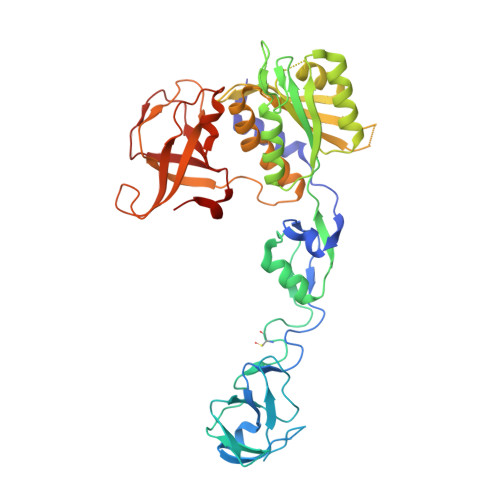
Insights into the Cnx1E catalyzed MPT-AMP hydrolysis.

Hercher, T.W.Krausze, J.Hoffmeister, S.Zwerschke, D.Lindel, T.Blankenfeldt, W.Mendel, R.R.Kruse, T.

(2020) Biosci Rep 40

  • DOI: https://doi.org/10.1042/BSR20191806
  • Primary Citation Related Structures: 
    6RMS

  • PubMed Abstract: 

    Molybdenum insertases (Mo-insertases) catalyze the final step of molybdenum cofactor (Moco) biosynthesis, an evolutionary old and highly conserved multi-step pathway. In the first step of the pathway, GTP serves as substrate for the formation of cyclic pyranopterin monophosphate, which is subsequently converted into molybdopterin (MPT) in the second pathway step. In the following synthesis steps, MPT is adenylated yielding MPT-AMP that is subsequently used as substrate for enzyme catalyzed molybdate insertion. Molybdate insertion and MPT-AMP hydrolysis are catalyzed by the Mo-insertase E-domain. Earlier work reported a highly conserved aspartate residue to be essential for Mo-insertase functionality. In this work, we confirmed the mechanistic relevance of this residue for the Arabidopsis thaliana Mo-insertase Cnx1E. We found that the conservative substitution of Cnx1E residue Asp274 by Glu (D274E) leads to an arrest of MPT-AMP hydrolysis and hence to the accumulation of MPT-AMP. We further showed that the MPT-AMP accumulation goes in hand with the accumulation of molybdate. By crystallization and structure determination of the Cnx1E variant D274E, we identified the potential reason for the missing hydrolysis activity in the disorder of the region spanning amino acids 269 to 274. We reasoned that this is caused by the inability of a glutamate in position 274 to coordinate the octahedral Mg2+-water complex in the Cnx1E active site.


  • Organizational Affiliation
    • TU Braunschweig, Institute of Plant Biology, Spielmannstrasse 7, 38106 Braunschweig, Germany.

Macromolecule Content 

  • Total Structure Weight: 46.02 kDa 
  • Atom Count: 3,217 
  • Modeled Residue Count: 397 
  • Deposited Residue Count: 427 
  • Unique protein chains: 1

Macromolecules

Find similar proteins by:|  3D Structure
Entity ID: 1
MoleculeChains  Sequence LengthOrganismDetailsImage
Molybdopterin biosynthesis protein CNX1427Arabidopsis thalianaMutation(s): 0 
Gene Names: CNX1At5g20990F22D1.6T10F18.20
EC: 2.10.1.1 (PDB Primary Data), 2.7.7.75 (PDB Primary Data)
UniProt
Find proteins for Q39054 (Arabidopsis thaliana)
Explore Q39054 
Go to UniProtKB:  Q39054
Entity Groups
Sequence Clusters30% Identity50% Identity70% Identity90% Identity95% Identity100% Identity
UniProt GroupQ39054
Sequence Annotations
Expand
Reference Sequence

Small Molecules

Ligands 5 Unique
IDChains Name / Formula / InChI Key2D Diagram3D Interactions
AMP
(Subject of Investigation/LOI)

Query on AMP


Download:Ideal Coordinates CCD File
E [auth A]ADENOSINE MONOPHOSPHATE
C10 H14 N5 O7 P
UDMBCSSLTHHNCD-KQYNXXCUSA-N
SO4

Query on SO4


Download:Ideal Coordinates CCD File
B [auth A]SULFATE ION
O4 S
QAOWNCQODCNURD-UHFFFAOYSA-L
IMD

Query on IMD


Download:Ideal Coordinates CCD File
D [auth A]IMIDAZOLE
C3 H5 N2
RAXXELZNTBOGNW-UHFFFAOYSA-O
EDO

Query on EDO


Download:Ideal Coordinates CCD File
C [auth A]1,2-ETHANEDIOL
C2 H6 O2
LYCAIKOWRPUZTN-UHFFFAOYSA-N
MG

Query on MG


Download:Ideal Coordinates CCD File
F [auth A]MAGNESIUM ION
Mg
JLVVSXFLKOJNIY-UHFFFAOYSA-N
Modified Residues  1 Unique
IDChains TypeFormula2D DiagramParent
CSX
Query on CSX
A
L-PEPTIDE LINKINGC3 H7 N O3 SCYS

Experimental Data & Validation

Experimental Data

  • Method: X-RAY DIFFRACTION
  • Resolution: 1.74 Å
  • R-Value Free:  0.220 (Depositor), 0.224 (DCC) 
  • R-Value Work:  0.201 (Depositor), 0.200 (DCC) 
  • R-Value Observed: 0.202 (Depositor) 
  • Method: SOLUTION SCATTERING
Space Group: I 2 2 2
Unit Cell:
Length ( Å )Angle ( ˚ )
a = 64.836α = 90
b = 119.48β = 90
c = 136.867γ = 90
Software Package:
Software NamePurpose
BUSTERrefinement
XDSdata reduction
autoPROCdata scaling
Aimlessdata scaling
Cootmodel building
BUSTERphasing

Structure Validation

View Full Validation Report


Ligand Structure Quality Assessment 


Entry History 

Deposition Data

  • Released Date: 2020-02-19 
  • Deposition Author(s): Krausze, J.

Revision History  (Full details and data files)

  • Version 1.0: 2020-02-19
    Type: Initial release
  • Version 1.1: 2024-01-24
    Changes: Data collection, Database references, Derived calculations, Refinement description