6RLO | pdb_00006rlo

Crystal structure of AT1412dm Fab fragment in complex with CD9 large extracellular loop


Experimental Data Snapshot

  • Method: X-RAY DIFFRACTION
  • Resolution: 2.20 Å
  • R-Value Free: 
    0.287 (Depositor), 0.268 (DCC) 
  • R-Value Work: 
    0.254 (Depositor) 
  • R-Value Observed: 
    0.255 (Depositor) 

Starting Model: experimental
View more details

wwPDB Validation 3D Report Full Report

Validation slider image for 6RLO

This is version 1.2 of the entry. See complete history

Literature

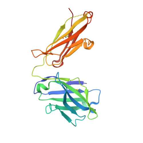
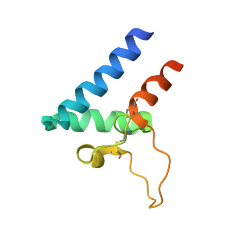
Structural basis of a homo-dimerization site in tetraspanin CD9 targeted by a melanoma patient-derived antibody

Neviani, V.Pos, W.Schotte, R.Wagner, K.Go, D.M.Fatmawati, C.Kedde, M.Claassen, Y.B.Kroon-Batenburg, L.Lutz, M.Verdegaal, E.M.E.van der Burg, S.H.Spits, H.Gros, P.

To be published.

Macromolecule Content 

  • Total Structure Weight: 233.99 kDa 
  • Atom Count: 15,875 
  • Modeled Residue Count: 2,027 
  • Deposited Residue Count: 2,132 
  • Unique protein chains: 3

Macromolecules

Find similar proteins by:|  3D Structure
Entity ID: 1
MoleculeChains  Sequence LengthOrganismDetailsImage
AT1412dm Fab Fragment (Heavy Chain)A,
D [auth C],
G [auth E],
J [auth G]
223Homo sapiensMutation(s): 0 
Find similar proteins by:|  3D Structure
Entity ID: 2
MoleculeChains  Sequence LengthOrganismDetailsImage
AT1412dm Fab Fragment (Light Chain)B,
E [auth D],
H [auth F],
K [auth H]
220Homo sapiensMutation(s): 0 
Find similar proteins by:|  3D Structure
Entity ID: 3
MoleculeChains  Sequence LengthOrganismDetailsImage
CD9 antigenC [auth I],
F [auth J],
I [auth K],
L
90Homo sapiensMutation(s): 0 
Gene Names: CD9MIC3TSPAN29GIG2
UniProt & NIH Common Fund Data Resources
Find proteins for P21926 (Homo sapiens)
Explore P21926 
Go to UniProtKB:  P21926
PHAROS:  P21926
GTEx:  ENSG00000010278 
Entity Groups
Sequence Clusters30% Identity50% Identity70% Identity90% Identity95% Identity100% Identity
UniProt GroupP21926
Sequence Annotations
Expand
Reference Sequence

Small Molecules

Ligands 3 Unique
IDChains Name / Formula / InChI Key2D Diagram3D Interactions
K

Query on K


Download:Ideal Coordinates CCD File
P [auth B]POTASSIUM ION
K
NPYPAHLBTDXSSS-UHFFFAOYSA-N
CL

Query on CL


Download:Ideal Coordinates CCD File
BA [auth F]
DA [auth G]
EA [auth H]
M [auth A]
N [auth A]
BA [auth F],
DA [auth G],
EA [auth H],
M [auth A],
N [auth A],
O [auth B],
R [auth I],
S [auth C],
T [auth C],
W [auth D],
X [auth D]
CHLORIDE ION
Cl
VEXZGXHMUGYJMC-UHFFFAOYSA-M
NA

Query on NA


Download:Ideal Coordinates CCD File
AA [auth D]
CA [auth F]
Q [auth B]
U [auth C]
V [auth C]
AA [auth D],
CA [auth F],
Q [auth B],
U [auth C],
V [auth C],
Y [auth D],
Z [auth D]
SODIUM ION
Na
FKNQFGJONOIPTF-UHFFFAOYSA-N

Experimental Data & Validation

Experimental Data

  • Method: X-RAY DIFFRACTION
  • Resolution: 2.20 Å
  • R-Value Free:  0.287 (Depositor), 0.268 (DCC) 
  • R-Value Work:  0.254 (Depositor) 
  • R-Value Observed: 0.255 (Depositor) 
Space Group: P 1
Unit Cell:
Length ( Å )Angle ( ˚ )
a = 73.776α = 71.12
b = 89.858β = 89.59
c = 91.533γ = 85.96
Software Package:
Software NamePurpose
DIALSdata reduction
Aimlessdata scaling
PHASERphasing
Cootmodel building
REFMACrefinement
PDB_EXTRACTdata extraction

Structure Validation

View Full Validation Report


Entry History 

& Funding Information

Deposition Data


Funding OrganizationLocationGrant Number
Netherlands Organisation for Scientific ResearchNetherlands024.002.009

Revision History  (Full details and data files)

  • Version 1.0: 2021-05-12
    Type: Initial release
  • Version 1.1: 2024-01-24
    Changes: Data collection, Database references, Refinement description
  • Version 1.2: 2024-10-09
    Changes: Structure summary