


Cryo-EM structure of TssA protein from Type VI secretion system of E. coli.
Nazarov, S., Demurtas, D., Shneider, M., Basler, M., Leiman, P.To be published.
Experimental Data Snapshot
Starting Model: experimental
View more details
wwPDB Validation 3D Report Full Report

Entity ID: 1 | |||||
|---|---|---|---|---|---|
| Molecule | Chains | Sequence Length | Organism | Details | Image |
| Putative type VI secretion protein | 532 | Escherichia coli | Mutation(s): 0 Gene Names: NCTC12950_00920 | 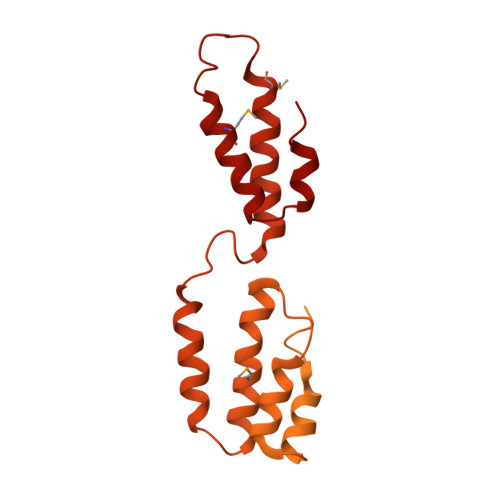 | |
| Modified Residues 1 Unique | |||||
|---|---|---|---|---|---|
| ID | Chains | Type | Formula | 2D Diagram | Parent |
| MSE Query on MSE | A | L-PEPTIDE LINKING | C5 H11 N O2 Se |  | MET |
| Task | Software Package | Version |
|---|---|---|
| RECONSTRUCTION | RELION | |
| MODEL REFINEMENT | PHENIX |
| Funding Organization | Location | Grant Number |
|---|---|---|
| Swiss National Science Foundation | Switzerland | BSSGI0_155778 |


