


Crystal structure of KDM3B in complex with 5-(1H-tetrazol-5-yl)quinolin-8-ol
Johansson, C., Newman, J.A., Kawamura, A., Schofield, C.J., Arrowsmith, C.H., Bountra, C., Edwards, A., Oppermann, U.C.T.To be published.
Experimental Data Snapshot
Starting Model: experimental
View more details
Entity ID: 1 | |||||
|---|---|---|---|---|---|
| Molecule | Chains | Sequence Length | Organism | Details | Image |
| Lysine-specific demethylase 3B | 351 | Homo sapiens | Mutation(s): 0 Gene Names: KDM3B, C5orf7, JHDM2B, JMJD1B, KIAA1082 EC: 1.14.11 (PDB Primary Data), 1.14.11.65 (UniProt) | 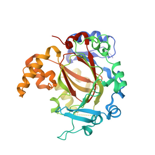 | |
UniProt & NIH Common Fund Data Resources | |||||
GTEx: ENSG00000120733 | |||||
Entity Groups | |||||
| Sequence Clusters | 30% Identity50% Identity70% Identity90% Identity95% Identity100% Identity | ||||
| UniProt Group | Q7LBC6 | ||||
Sequence AnnotationsExpand | |||||
Reference Sequence | |||||
| Ligands 4 Unique | |||||
|---|---|---|---|---|---|
| ID | Chains | Name / Formula / InChI Key | 2D Diagram | 3D Interactions | |
| JX8 (Subject of Investigation/LOI) Download:Ideal Coordinates CCD File | E [auth A], K [auth B] | 5-(1~{H}-1,2,3,4-tetrazol-5-yl)quinolin-8-ol C10 H7 N5 O COSOIVLLWHGBPW-UHFFFAOYSA-N |  | ||
| EDO Download:Ideal Coordinates CCD File | C [auth A], D [auth A], H [auth B], I [auth B], J [auth B] | 1,2-ETHANEDIOL C2 H6 O2 LYCAIKOWRPUZTN-UHFFFAOYSA-N |  | ||
| MN Download:Ideal Coordinates CCD File | F [auth A], L [auth B] | MANGANESE (II) ION Mn WAEMQWOKJMHJLA-UHFFFAOYSA-N |  | ||
| CL Download:Ideal Coordinates CCD File | G [auth A] | CHLORIDE ION Cl VEXZGXHMUGYJMC-UHFFFAOYSA-M |  | ||
| Length ( Å ) | Angle ( ˚ ) |
|---|---|
| a = 56.929 | α = 90 |
| b = 93.754 | β = 107.29 |
| c = 90.494 | γ = 90 |
| Software Name | Purpose |
|---|---|
| XDS | data reduction |
| Aimless | data scaling |
| PHASER | phasing |
| PHENIX | refinement |
| PDB_EXTRACT | data extraction |


