6RBC | pdb_00006rbc

X-ray structure of human glutamate carboxypeptidase II (GCPII) in complex with a inhibitor KB1157


Experimental Data Snapshot

  • Method: X-RAY DIFFRACTION
  • Resolution: 1.77 Å
  • R-Value Free: 
    0.191 (Depositor), 0.195 (DCC) 
  • R-Value Work: 
    0.164 (Depositor), 0.169 (DCC) 
  • R-Value Observed: 
    0.165 (Depositor) 

Starting Model: experimental
View more details

wwPDB Validation 3D Report Full Report

Validation slider image for 6RBC

Ligand Structure Quality Assessment 


This is version 2.3 of the entry. See complete history

Literature

Novel beta- and gamma-Amino Acid-Derived Inhibitors of Prostate-Specific Membrane Antigen.

Kim, K.Kwon, H.Barinka, C.Motlova, L.Nam, S.Choi, D.Ha, H.Nam, H.Son, S.H.Minn, I.Pomper, M.G.Yang, X.Kutil, Z.Byun, Y.

(2020) J Med Chem 63: 3261-3273

  • DOI: https://doi.org/10.1021/acs.jmedchem.9b02022
  • Primary Citation Related Structures: 
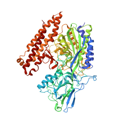
    6RBC, 6S1X

  • PubMed Abstract: 

    Prostate-specific membrane antigen (PSMA) is an excellent biomarker for the early diagnosis of prostate cancer progression and metastasis. The most promising PSMA-targeted agents in the clinical phase are based on the Lys-urea-Glu motif, in which Lys and Glu are α-(l)-amino acids. In this study, we aimed to determine the effect of β- and γ-amino acids in the S1 pocket on the binding affinity for PSMA. We synthesized and evaluated the β- and γ-amino acid analogues with ( S )- or ( R )-configuration with keeping α-(l)-Glu as the S1'-binding pharmacophore. The structure-activity relationship studies identified that compound 13c , a β-amino acid analogue with ( R )-configuration, exhibited the most potent PSMA inhibitory activity with an IC 50 value of 3.97 nM. The X-ray crystal structure of PSMA in complex with 13c provided a mechanistic basis for the stereochemical preference of PSMA, which can guide the development of future PSMA inhibitors.


  • Organizational Affiliation
    • College of Pharmacy, Korea University, 2511 Sejong-ro, Jochiwon-eup, Sejong 30019, Republic of Korea.

Macromolecule Content 

  • Total Structure Weight: 82.9 kDa 
  • Atom Count: 6,603 
  • Modeled Residue Count: 694 
  • Deposited Residue Count: 707 
  • Unique protein chains: 1

Macromolecules

Find similar proteins by:|  3D Structure
Entity ID: 1
MoleculeChains  Sequence LengthOrganismDetailsImage
Glutamate carboxypeptidase 2707Homo sapiensMutation(s): 0 
Gene Names: FOLH1FOLHNAALAD1PSMPSMAGIG27
EC: 3.4.17.21
UniProt & NIH Common Fund Data Resources
Find proteins for Q04609 (Homo sapiens)
Explore Q04609 
Go to UniProtKB:  Q04609
PHAROS:  Q04609
GTEx:  ENSG00000086205 
Entity Groups
Sequence Clusters30% Identity50% Identity70% Identity90% Identity95% Identity100% Identity
UniProt GroupQ04609
Glycosylation
Glycosylation Sites: 7Go to GlyGen: Q04609-1
Sequence Annotations
Expand
Reference Sequence

Oligosaccharides

Help  
Entity ID: 2
MoleculeChains Length2D Diagram GlycosylationD Interactions
2-acetamido-2-deoxy-beta-D-glucopyranose-(1-4)-2-acetamido-2-deoxy-beta-D-glucopyranose
B, C
2N-Glycosylation
Glycosylation Resources
GlyTouCan: G42666HT
GlyCosmos: G42666HT
GlyGen: G42666HT
Entity ID: 3
MoleculeChains Length2D Diagram GlycosylationD Interactions
alpha-D-mannopyranose-(1-3)-beta-D-mannopyranose-(1-4)-2-acetamido-2-deoxy-beta-D-glucopyranose-(1-4)-2-acetamido-2-deoxy-beta-D-glucopyranose
D
4N-Glycosylation
Glycosylation Resources
GlyTouCan: G81315DD
GlyCosmos: G81315DD
GlyGen: G81315DD

Small Molecules

Ligands 5 Unique
IDChains Name / Formula / InChI Key2D Diagram3D Interactions
JX5

Query on JX5


Download:Ideal Coordinates CCD File
M [auth A](2~{S})-2-[[(2~{R})-2-[4-[(4-iodophenyl)carbonylamino]butanoylamino]-3-oxidanyl-3-oxidanylidene-propyl]carbamoylamino]pentanedioic acid
C20 H25 I N4 O9
AOGSPOQTLUVJFT-UONOGXRCSA-N
NAG

Query on NAG


Download:Ideal Coordinates CCD File
E [auth A],
F [auth A],
G [auth A],
H [auth A]
2-acetamido-2-deoxy-beta-D-glucopyranose
C8 H15 N O6
OVRNDRQMDRJTHS-FMDGEEDCSA-N
ZN

Query on ZN


Download:Ideal Coordinates CCD File
I [auth A],
J [auth A]
ZINC ION
Zn
PTFCDOFLOPIGGS-UHFFFAOYSA-N
CA

Query on CA


Download:Ideal Coordinates CCD File
K [auth A]CALCIUM ION
Ca
BHPQYMZQTOCNFJ-UHFFFAOYSA-N
CL

Query on CL


Download:Ideal Coordinates CCD File
L [auth A]CHLORIDE ION
Cl
VEXZGXHMUGYJMC-UHFFFAOYSA-M
Binding Affinity Annotations 
IDSourceBinding Affinity
JX5 BindingDB:  6RBC IC50: 4 (nM) from 1 assay(s)

Experimental Data & Validation

Experimental Data

  • Method: X-RAY DIFFRACTION
  • Resolution: 1.77 Å
  • R-Value Free:  0.191 (Depositor), 0.195 (DCC) 
  • R-Value Work:  0.164 (Depositor), 0.169 (DCC) 
  • R-Value Observed: 0.165 (Depositor) 
Space Group: I 2 2 2
Unit Cell:
Length ( Å )Angle ( ˚ )
a = 101.551α = 90
b = 130.47β = 90
c = 159.045γ = 90
Software Package:
Software NamePurpose
REFMACrefinement
XDSdata reduction
Aimlessdata scaling
REFMACphasing

Structure Validation

View Full Validation Report


Ligand Structure Quality Assessment 


Entry History 

Deposition Data

Revision History  (Full details and data files)

  • Version 1.0: 2020-05-13
    Type: Initial release
  • Version 2.0: 2020-07-29
    Type: Remediation
    Reason: Carbohydrate remediation
    Changes: Atomic model, Data collection, Derived calculations, Structure summary
  • Version 2.1: 2020-11-25
    Changes: Database references, Derived calculations, Structure summary
  • Version 2.2: 2024-01-24
    Changes: Data collection, Database references, Refinement description
  • Version 2.3: 2024-11-13
    Changes: Structure summary