6R7B | pdb_00006r7b

Crystal structure of Csx1 in complex with cyclic oligoadenylate cOA4 conformation 1


Experimental Data Snapshot

  • Method: X-RAY DIFFRACTION
  • Resolution: 3.12 Å
  • R-Value Free: 
    0.229 (Depositor), 0.247 (DCC) 
  • R-Value Work: 
    0.180 (Depositor) 
  • R-Value Observed: 
    0.183 (Depositor) 

Starting Model: experimental
View more details

wwPDB Validation 3D Report Full Report

Validation slider image for 6R7B

This is version 1.2 of the entry. See complete history

Literature

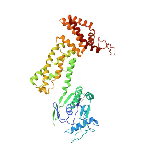
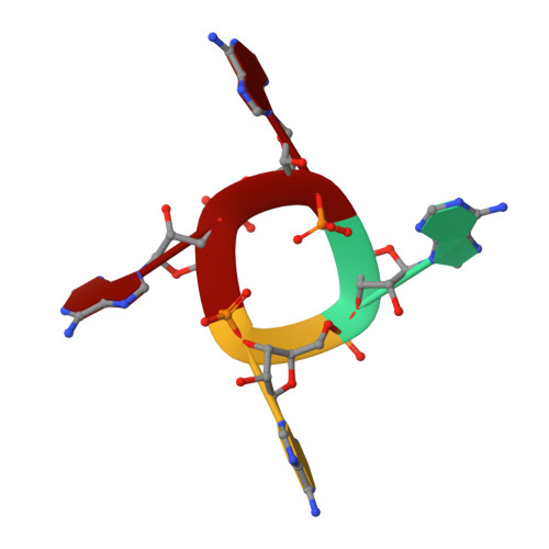
Structure of Csx1-cOA4complex reveals the basis of RNA decay in Type III-B CRISPR-Cas.

Molina, R.Stella, S.Feng, M.Sofos, N.Jauniskis, V.Pozdnyakova, I.Lopez-Mendez, B.She, Q.Montoya, G.

(2019) Nat Commun 10: 4302-4302

  • DOI: https://doi.org/10.1038/s41467-019-12244-z
  • Primary Citation Related Structures: 
    6QZQ, 6QZT, 6R7B, 6R9R

  • PubMed Abstract: 

    Type III CRISPR-Cas multisubunit complexes cleave ssRNA and ssDNA. These activities promote the generation of cyclic oligoadenylate (cOA), which activates associated CRISPR-Cas RNases from the Csm/Csx families, triggering a massive RNA decay to provide immunity from genetic invaders. Here we present the structure of Sulfolobus islandicus (Sis) Csx1-cOA 4 complex revealing the allosteric activation of its RNase activity. SisCsx1 is a hexamer built by a trimer of dimers. Each dimer forms a cOA 4 binding site and a ssRNA catalytic pocket. cOA 4 undergoes a conformational change upon binding in the second messenger binding site activating ssRNA degradation in the catalytic pockets. Activation is transmitted in an allosteric manner through an intermediate HTH domain, which joins the cOA 4 and catalytic sites. The RNase functions in a sequential cooperative fashion, hydrolyzing phosphodiester bonds in 5'-C-C-3'. The degradation of cOA 4 by Ring nucleases deactivates SisCsx1, suggesting that this enzyme could be employed in biotechnological applications.


  • Organizational Affiliation
    • Structural Molecular Biology Group, Novo Nordisk Foundation Centre for Protein Research, Faculty of Health and Medical Sciences University of Copenhagen, Blegdamsvej 3-B, Copenhagen, 2200, Denmark.

Macromolecule Content 

  • Total Structure Weight: 158.12 kDa 
  • Atom Count: 11,153 
  • Modeled Residue Count: 1,368 
  • Deposited Residue Count: 1,370 
  • Unique protein chains: 1
  • Unique nucleic acid chains: 1

Macromolecules


Find similar proteins by:|  3D Structure
Entity ID: 1
MoleculeChains  Sequence LengthOrganismDetailsImage
CRISPR-associated (Cas) DxTHG family
A, B, C
454Saccharolobus islandicusMutation(s): 0 
Gene Names: SiRe_0884
UniProt
Find proteins for F0NE21 (Saccharolobus islandicus (strain REY15A))
Explore F0NE21 
Go to UniProtKB:  F0NE21
Entity Groups
Sequence Clusters30% Identity50% Identity70% Identity90% Identity95% Identity100% Identity
UniProt GroupF0NE21
Sequence Annotations
Expand
Reference Sequence
Find similar nucleic acids by:  Sequence
Entity ID: 2
MoleculeChains LengthOrganismImage
RNA (5'-R(P*AP*AP*AP*A)-3')
D, E
4synthetic construct
Sequence Annotations
Expand
Reference Sequence

Experimental Data & Validation

Experimental Data

  • Method: X-RAY DIFFRACTION
  • Resolution: 3.12 Å
  • R-Value Free:  0.229 (Depositor), 0.247 (DCC) 
  • R-Value Work:  0.180 (Depositor) 
  • R-Value Observed: 0.183 (Depositor) 
Space Group: I 21 21 21
Unit Cell:
Length ( Å )Angle ( ˚ )
a = 107.08α = 90
b = 117.68β = 90
c = 357.26γ = 90
Software Package:
Software NamePurpose
BUSTERrefinement
XDSdata reduction
Aimlessdata scaling
PHASERphasing

Structure Validation

View Full Validation Report


Entry History 

& Funding Information

Deposition Data


Funding OrganizationLocationGrant Number
Novo Nordisk FoundationDenmarkNNF14CC0001

Revision History  (Full details and data files)

  • Version 1.0: 2019-10-02
    Type: Initial release
  • Version 1.1: 2024-01-24
    Changes: Data collection, Database references, Refinement description
  • Version 1.2: 2024-10-16
    Changes: Structure summary