6R57

Crystal structure of PPEP-1(E143A/Y178F/E184A) in complex with substrate peptide Ac-EVNPPVP-CONH2


Experimental Data Snapshot

  • Method: X-RAY DIFFRACTION
  • Resolution: 1.90 Å
  • R-Value Free: 0.187 
  • R-Value Work: 0.160 
  • R-Value Observed: 0.161 

Starting Model: experimental
View more details

wwPDB Validation   3D Report Full Report


This is version 1.3 of the entry. See complete history


Literature

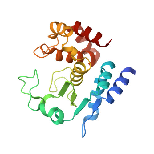
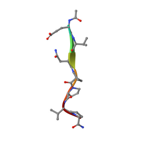
Molecular determinants of the mechanism and substrate specificity ofClostridium difficileproline-proline endopeptidase-1.

Pichlo, C.Juetten, L.Wojtalla, F.Schacherl, M.Diaz, D.Baumann, U.

(2019) J Biol Chem 294: 11525-11535

  • DOI: https://doi.org/10.1074/jbc.RA119.009029
  • Primary Citation of Related Structures:  
    6R4W, 6R4X, 6R4Y, 6R4Z, 6R50, 6R51, 6R52, 6R53, 6R54, 6R55, 6R56, 6R57, 6R58, 6R59, 6R5A, 6R5B, 6R5C, 6R9Z

  • PubMed Abstract: 

    Pro-Pro endopeptidase-1 (PPEP-1) is a secreted metalloprotease from the bacterial pathogen Clostridium difficile that cleaves two endogenous adhesion proteins. PPEP-1 is therefore important for bacterial motility and hence for efficient gut colonization during infection. PPEP-1 exhibits a unique specificity for Pro-Pro peptide bonds within the consensus sequence VNP↓PVP. In this study, we combined information from crystal and NMR structures with mutagenesis and enzyme kinetics to investigate the mechanism and substrate specificity of PPEP-1. Our analyses revealed that the substrate-binding cleft of PPEP-1 is shaped complementarily to the major conformation of the substrate in solution. We found that it possesses features that accept a tertiary amide and help discriminate P1' residues by their amide hydrogen bond-donating potential. We also noted that residues Lys-101, Trp-103, and Glu-184 are crucial for proteolytic activity. Upon substrate binding, these residues position a flexible loop over the substrate-binding cleft and modulate the second coordination sphere of the catalytic zinc ion. On the basis of these findings, we propose an induced-fit model in which prestructured substrates are recognized followed by substrate positioning within the active-site cleft and a concomitant increase in the Lewis acidity of the catalytic Zn 2+ ion. In conclusion, our findings provide detailed structural and mechanistic insights into the substrate recognition and specificity of PPEP-1 from the common gut pathogen C. difficile .


  • Organizational Affiliation

    Department of Chemistry, Institute of Biochemistry, University of Cologne, 50674 Cologne, Germany.


Macromolecules
Find similar proteins by:  (by identity cutoff)  |  3D Structure
Entity ID: 1
MoleculeChains Sequence LengthOrganismDetailsImage
Pro-Pro endopeptidase
A, B
198Clostridioides difficileMutation(s): 3 
Gene Names: zmp1ppep-1CD630_28300
EC: 3.4.24.89
UniProt
Find proteins for Q183R7 (Clostridioides difficile (strain 630))
Explore Q183R7 
Go to UniProtKB:  Q183R7
Entity Groups  
Sequence Clusters30% Identity50% Identity70% Identity90% Identity95% Identity100% Identity
UniProt GroupQ183R7
Sequence Annotations
Expand
  • Reference Sequence

Find similar proteins by:  Sequence   |   3D Structure  

Entity ID: 2
MoleculeChains Sequence LengthOrganismDetailsImage
ACE-GLU-VAL-ASN-PRO-PRO-VAL-LPD
C, D
8Clostridioides difficileMutation(s): 0 
Sequence Annotations
Expand
  • Reference Sequence
Small Molecules
Ligands 1 Unique
IDChains Name / Formula / InChI Key2D Diagram3D Interactions
ZN
Query on ZN

Download Ideal Coordinates CCD File 
E [auth B]ZINC ION
Zn
PTFCDOFLOPIGGS-UHFFFAOYSA-N
Modified Residues  1 Unique
IDChains TypeFormula2D DiagramParent
LPD
Query on LPD
C, D
L-PEPTIDE LINKINGC5 H10 N2 OPRO
Experimental Data & Validation

Experimental Data

  • Method: X-RAY DIFFRACTION
  • Resolution: 1.90 Å
  • R-Value Free: 0.187 
  • R-Value Work: 0.160 
  • R-Value Observed: 0.161 
  • Space Group: P 1 21 1
Unit Cell:
Length ( Å )Angle ( ˚ )
a = 37.202α = 90
b = 43.005β = 96.71
c = 121.908γ = 90
Software Package:
Software NamePurpose
PHENIXrefinement
XDSdata reduction
XDSdata scaling
PHENIXphasing

Structure Validation

View Full Validation Report


Entry History 

Deposition Data

Revision History  (Full details and data files)

  • Version 1.0: 2019-06-12
    Type: Initial release
  • Version 1.1: 2019-06-26
    Changes: Data collection, Database references
  • Version 1.2: 2019-08-07
    Changes: Data collection, Database references
  • Version 1.3: 2024-01-24
    Changes: Data collection, Database references, Derived calculations, Refinement description