6R3V | pdb_00006r3v

Crystal Structure of RhoA-GDP-Pi in Complex with RhoGAP


Experimental Data Snapshot

  • Method: X-RAY DIFFRACTION
  • Resolution: 1.75 Å
  • R-Value Free: 
    0.207 (Depositor), 0.210 (DCC) 
  • R-Value Work: 
    0.177 (Depositor), 0.190 (DCC) 
  • R-Value Observed: 
    0.178 (Depositor) 

Starting Model: experimental
View more details

wwPDB Validation   3D Report Full Report


Ligand Structure Quality Assessment 

Created with Raphaël 2.3.0Worse 01 BetterLigand structure goodness of fit to experimental dataBest fitted GDPClick on this verticalbar to view detailsBest fitted PO4Click on this verticalbar to view details

This is version 1.4 of the entry. See complete history


Literature

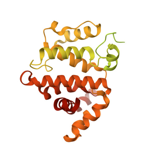
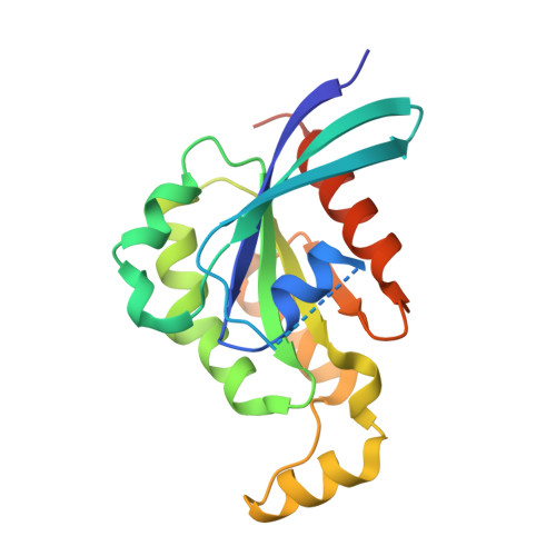
A GAP-GTPase-GDP-PiIntermediate Crystal Structure Analyzed by DFT Shows GTP Hydrolysis Involves Serial Proton Transfers.

Molt Jr., R.W.Pellegrini, E.Jin, Y.

(2019) Chemistry 25: 8484-8488

  • DOI: https://doi.org/10.1002/chem.201901627
  • Primary Citation of Related Structures:  
    6R3V

  • PubMed Abstract: 

    Cell signaling by small G proteins uses an ON to OFF signal based on conformational changes following the hydrolysis of GTP to GDP and release of dihydrogen phosphate (P i ). The catalytic mechanism of GTP hydrolysis by RhoA is strongly accelerated by a GAP protein and is now well defined, but timing of inorganic phosphate release and signal change remains unresolved. We have generated a quaternary complex for RhoA-GAP-GDP-P i . Its 1.75 Å crystal structure shows geometry for ionic and hydrogen bond coordination of GDP and P i in an intermediate state. It enables the selection of a QM core for DFT exploration of a 20 H-bonded network. This identifies serial locations of the two mobile protons from the original nucleophilic water molecule, showing how they move in three rational steps to form a stable quaternary complex. It also suggests how two additional proton transfer steps can facilitate P i release.


  • Organizational Affiliation

    Department of Biochemistry & Molecular Biology, Indiana University School of Medicine, Indianapolis, Indiana, 46202, USA.


Macromolecules
Find similar proteins by:  (by identity cutoff)  |  3D Structure
Entity ID: 1
MoleculeChains Sequence LengthOrganismDetailsImage
Rho GTPase-activating protein 1439Homo sapiensMutation(s): 0 
Gene Names: ARHGAP1CDC42GAPRHOGAP1
UniProt & NIH Common Fund Data Resources
Find proteins for Q07960 (Homo sapiens)
Explore Q07960 
Go to UniProtKB:  Q07960
PHAROS:  Q07960
GTEx:  ENSG00000175220 
Entity Groups  
Sequence Clusters30% Identity50% Identity70% Identity90% Identity95% Identity100% Identity
UniProt GroupQ07960
Sequence Annotations
Expand
  • Reference Sequence
Find similar proteins by:  (by identity cutoff)  |  3D Structure
Entity ID: 2
MoleculeChains Sequence LengthOrganismDetailsImage
Transforming protein RhoA193Homo sapiensMutation(s): 0 
Gene Names: RHOAARH12ARHARHO12
EC: 3.6.5.2
UniProt & NIH Common Fund Data Resources
Find proteins for P61586 (Homo sapiens)
Explore P61586 
Go to UniProtKB:  P61586
PHAROS:  P61586
GTEx:  ENSG00000067560 
Entity Groups  
Sequence Clusters30% Identity50% Identity70% Identity90% Identity95% Identity100% Identity
UniProt GroupP61586
Sequence Annotations
Expand
  • Reference Sequence
Small Molecules
Ligands 5 Unique
IDChains Name / Formula / InChI Key2D Diagram3D Interactions
GDP (Subject of Investigation/LOI)
Query on GDP

Download Ideal Coordinates CCD File 
E [auth B]GUANOSINE-5'-DIPHOSPHATE
C10 H15 N5 O11 P2
QGWNDRXFNXRZMB-UUOKFMHZSA-N
MES
Query on MES

Download Ideal Coordinates CCD File 
D [auth B]2-(N-MORPHOLINO)-ETHANESULFONIC ACID
C6 H13 N O4 S
SXGZJKUKBWWHRA-UHFFFAOYSA-N
DTT
Query on DTT

Download Ideal Coordinates CCD File 
H [auth B]2,3-DIHYDROXY-1,4-DITHIOBUTANE
C4 H10 O2 S2
VHJLVAABSRFDPM-IMJSIDKUSA-N
PO4 (Subject of Investigation/LOI)
Query on PO4

Download Ideal Coordinates CCD File 
C [auth A],
G [auth B]
PHOSPHATE ION
O4 P
NBIIXXVUZAFLBC-UHFFFAOYSA-K
MG (Subject of Investigation/LOI)
Query on MG

Download Ideal Coordinates CCD File 
F [auth B]MAGNESIUM ION
Mg
JLVVSXFLKOJNIY-UHFFFAOYSA-N
Experimental Data & Validation

Experimental Data

  • Method: X-RAY DIFFRACTION
  • Resolution: 1.75 Å
  • R-Value Free:  0.207 (Depositor), 0.210 (DCC) 
  • R-Value Work:  0.177 (Depositor), 0.190 (DCC) 
  • R-Value Observed: 0.178 (Depositor) 
Space Group: C 2 2 21
Unit Cell:
Length ( Å )Angle ( ˚ )
a = 114.06α = 90
b = 122.78β = 90
c = 67.19γ = 90
Software Package:
Software NamePurpose
REFMACrefinement
XDSdata reduction
Aimlessdata scaling
MOLREPphasing

Structure Validation

View Full Validation Report


Ligand Structure Quality Assessment 

Created with Raphaël 2.3.0Worse 01 BetterLigand structure goodness of fit to experimental dataBest fitted GDPClick on this verticalbar to view detailsBest fitted PO4Click on this verticalbar to view details

Entry History & Funding Information

Deposition Data

  • Released Date: 2019-05-08 
  • Deposition Author(s): Jin, Y.

Funding OrganizationLocationGrant Number
Wellcome TrustUnited Kingdom209057/Z/17/Z

Revision History  (Full details and data files)

  • Version 1.0: 2019-05-08
    Type: Initial release
  • Version 1.1: 2019-07-10
    Changes: Data collection
  • Version 1.2: 2019-07-17
    Changes: Data collection, Database references
  • Version 1.3: 2024-01-24
    Changes: Data collection, Database references, Derived calculations, Refinement description
  • Version 1.4: 2024-11-06
    Changes: Structure summary