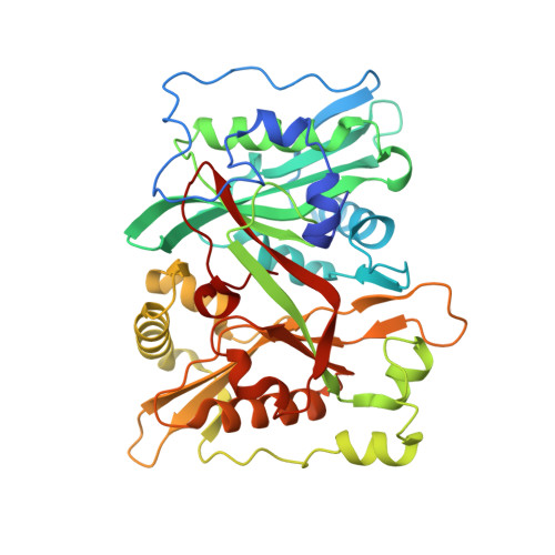
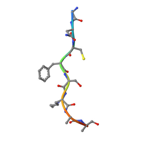
6QRM | pdb_00006qrm

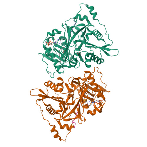
HsNMT1 in complex with both MyrCoA and GNCFSKRRAA substrates


Experimental Data Snapshot

  • Method: X-RAY DIFFRACTION
  • Resolution: 2.30 Å
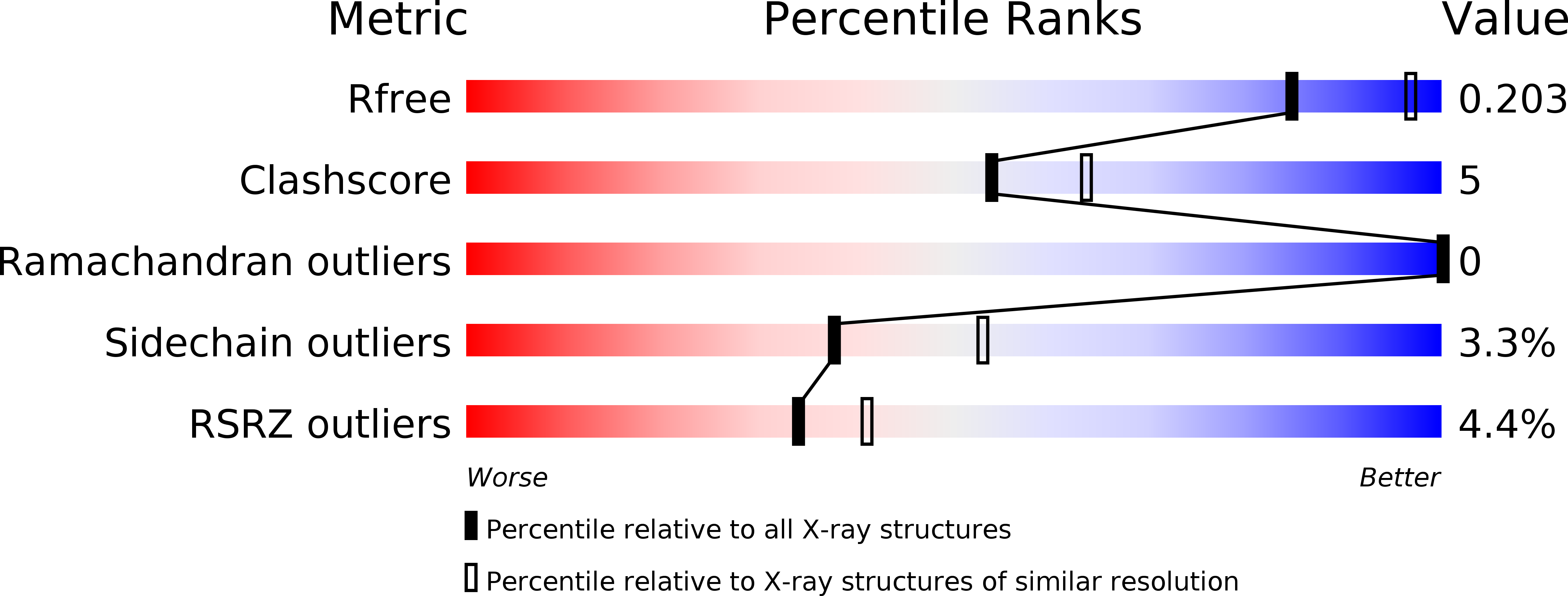
  • R-Value Free: 
    0.203 (Depositor), 0.200 (DCC) 
  • R-Value Work: 
    0.172 (Depositor), 0.170 (DCC) 
  • R-Value Observed: 
    0.174 (Depositor) 

Starting Model: experimental
View more details

wwPDB Validation   3D Report Full Report


Ligand Structure Quality Assessment 

Created with Raphaël 2.3.0Worse 01 BetterLigand structure goodness of fit to experimental dataBest fitted MYAClick on this verticalbar to view details

This is version 1.2 of the entry. See complete history


Literature

High-resolution snapshots of human N-myristoyltransferase in action illuminate a mechanism promoting N-terminal Lys and Gly myristoylation.

Dian, C.Perez-Dorado, I.Riviere, F.Asensio, T.Legrand, P.Ritzefeld, M.Shen, M.Cota, E.Meinnel, T.Tate, E.W.Giglione, C.

(2020) Nat Commun 11: 1132-1132

  • DOI: https://doi.org/10.1038/s41467-020-14847-3
  • Primary Citation of Related Structures:  
    6EHJ, 6QRM, 6SJZ, 6SK2, 6SK3, 6SK8, 6SKJ

  • PubMed Abstract: 

    The promising drug target N-myristoyltransferase (NMT) catalyses an essential protein modification thought to occur exclusively at N-terminal glycines (Gly). Here, we present high-resolution human NMT1 structures co-crystallised with reactive cognate lipid and peptide substrates, revealing high-resolution snapshots of the entire catalytic mechanism from the initial to final reaction states. Structural comparisons, together with biochemical analysis, provide unforeseen details about how NMT1 reaches a catalytically competent conformation in which the reactive groups are brought into close proximity to enable catalysis. We demonstrate that this mechanism further supports efficient and unprecedented myristoylation of an N-terminal lysine side chain, providing evidence that NMT acts both as N-terminal-lysine and glycine myristoyltransferase.


  • Organizational Affiliation

    Université Paris-Saclay, CEA, CNRS, Institute for Integrative Biology of the Cell (I2BC), Gif-sur-Yvette, 91198, France.


Macromolecules
Find similar proteins by:  (by identity cutoff)  |  3D Structure
Entity ID: 1
MoleculeChains Sequence LengthOrganismDetailsImage
Glycylpeptide N-tetradecanoyltransferase 1
A, B
402Homo sapiensMutation(s): 0 
Gene Names: NMT1NMT
EC: 2.3.1.97 (PDB Primary Data), 2.3.1 (UniProt)
UniProt & NIH Common Fund Data Resources
Find proteins for P30419 (Homo sapiens)
Explore P30419 
Go to UniProtKB:  P30419
PHAROS:  P30419
GTEx:  ENSG00000136448 
Entity Groups  
Sequence Clusters30% Identity50% Identity70% Identity90% Identity95% Identity100% Identity
UniProt GroupP30419
Sequence Annotations
Expand
  • Reference Sequence

Find similar proteins by:  Sequence   |   3D Structure  

Entity ID: 2
MoleculeChains Sequence LengthOrganismDetailsImage
Apoptosis-inducing factor 3
C, D
10Homo sapiensMutation(s): 2 
EC: 1
UniProt & NIH Common Fund Data Resources
Find proteins for Q96NN9 (Homo sapiens)
Explore Q96NN9 
Go to UniProtKB:  Q96NN9
PHAROS:  Q96NN9
GTEx:  ENSG00000183773 
Entity Groups  
Sequence Clusters30% Identity50% Identity70% Identity90% Identity95% Identity100% Identity
UniProt GroupQ96NN9
Sequence Annotations
Expand
  • Reference Sequence
Small Molecules
Ligands 5 Unique
IDChains Name / Formula / InChI Key2D Diagram3D Interactions
MYA (Subject of Investigation/LOI)
Query on MYA

Download Ideal Coordinates CCD File 
I [auth B]TETRADECANOYL-COA
C35 H62 N7 O17 P3 S
DUAFKXOFBZQTQE-QSGBVPJFSA-N
COA
Query on COA

Download Ideal Coordinates CCD File 
E [auth A]COENZYME A
C21 H36 N7 O16 P3 S
RGJOEKWQDUBAIZ-IBOSZNHHSA-N
MYR
Query on MYR

Download Ideal Coordinates CCD File 
M [auth C]MYRISTIC ACID
C14 H28 O2
TUNFSRHWOTWDNC-UHFFFAOYSA-N
GOL
Query on GOL

Download Ideal Coordinates CCD File 
F [auth A],
G [auth A],
J [auth B],
K [auth B]
GLYCEROL
C3 H8 O3
PEDCQBHIVMGVHV-UHFFFAOYSA-N
CL
Query on CL

Download Ideal Coordinates CCD File 
H [auth A],
L [auth B]
CHLORIDE ION
Cl
VEXZGXHMUGYJMC-UHFFFAOYSA-M
Experimental Data & Validation

Experimental Data

  • Method: X-RAY DIFFRACTION
  • Resolution: 2.30 Å
  • R-Value Free:  0.203 (Depositor), 0.200 (DCC) 
  • R-Value Work:  0.172 (Depositor), 0.170 (DCC) 
  • R-Value Observed: 0.174 (Depositor) 
Space Group: P 21 21 2
Unit Cell:
Length ( Å )Angle ( ˚ )
a = 79.437α = 90
b = 178.931β = 90
c = 58.412γ = 90
Software Package:
Software NamePurpose
PHENIXrefinement
XDSdata reduction
Aimlessdata scaling
MOLREPphasing

Structure Validation

View Full Validation Report


Ligand Structure Quality Assessment 

Created with Raphaël 2.3.0Worse 01 BetterLigand structure goodness of fit to experimental dataBest fitted MYAClick on this verticalbar to view details

Entry History & Funding Information

Deposition Data


Funding OrganizationLocationGrant Number
French National Research AgencyFranceANR-2010-BLAN-1611-01
French National Research AgencyFranceANR-10-LABX-0040-SPS

Revision History  (Full details and data files)

  • Version 1.0: 2020-03-18
    Type: Initial release
  • Version 1.1: 2024-01-24
    Changes: Data collection, Database references, Refinement description
  • Version 1.2: 2024-11-20
    Changes: Structure summary