6QQ3

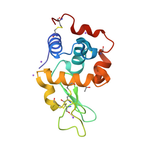
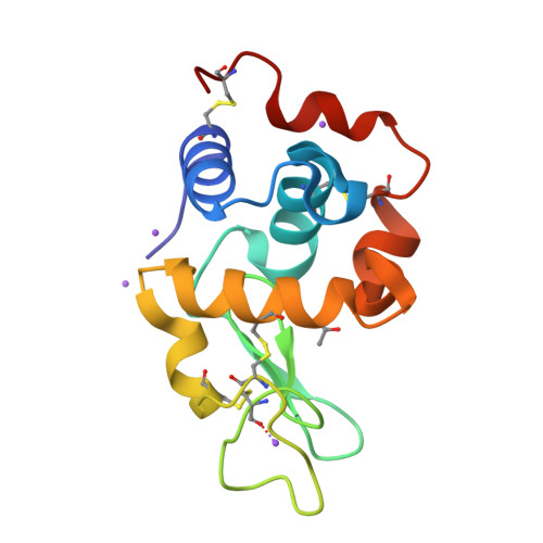
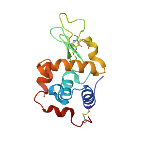
The room temperature structure of lysozyme via the acoustic levitation of a droplet


Experimental Data Snapshot

  • Method: X-RAY DIFFRACTION
  • Resolution: 1.53 Å
  • R-Value Free: 0.201 
  • R-Value Work: 0.177 
  • R-Value Observed: 0.178 

Starting Model: experimental
View more details

wwPDB Validation   3D Report Full Report


This is version 1.4 of the entry. See complete history


Literature

Non-Contact Universal Sample Presentation for Room Temperature Macromolecular Crystallography Using Acoustic Levitation.

Morris, R.H.Dye, E.R.Axford, D.Newton, M.I.Beale, J.H.Docker, P.T.

(2019) Sci Rep 9: 12431-12431

  • DOI: https://doi.org/10.1038/s41598-019-48612-4
  • Primary Citation of Related Structures:  
    6QQ3

  • PubMed Abstract: 

    Macromolecular Crystallography is a powerful and valuable technique to assess protein structures. Samples are commonly cryogenically cooled to minimise radiation damage effects from the X-ray beam, but low temperatures hinder normal protein functions and this procedure can introduce structural artefacts. Previous experiments utilising acoustic levitation for beamline science have focused on Langevin horns which deliver significant power to the confined droplet and are complex to set up accurately. In this work, the low power, portable TinyLev acoustic levitation system is used in combination with an approach to dispense and contain droplets, free of physical sample support to aid protein crystallography experiments. This method facilitates efficient X-ray data acquisition in ambient conditions compatible with dynamic studies. Levitated samples remain free of interference from fixed sample mounts, receive negligible heating, do not suffer significant evaporation and since the system occupies a small volume, can be readily installed at other light sources.


  • Organizational Affiliation

    School of Science and Technology, Nottingham Trent University, Nottingham, NG11 8NS, UK. rob.morris@ntu.ac.uk.


Macromolecules
Find similar proteins by:  (by identity cutoff)  |  3D Structure
Entity ID: 1
MoleculeChains Sequence LengthOrganismDetailsImage
Lysozyme CA [auth B]129Gallus gallusMutation(s): 0 
Gene Names: LYZ
EC: 3.2.1.17
UniProt
Find proteins for P00698 (Gallus gallus)
Explore P00698 
Go to UniProtKB:  P00698
Entity Groups  
Sequence Clusters30% Identity50% Identity70% Identity90% Identity95% Identity100% Identity
UniProt GroupP00698
Sequence Annotations
Expand
  • Reference Sequence
Small Molecules
Ligands 3 Unique
IDChains Name / Formula / InChI Key2D Diagram3D Interactions
ACT
Query on ACT

Download Ideal Coordinates CCD File 
C [auth B]ACETATE ION
C2 H3 O2
QTBSBXVTEAMEQO-UHFFFAOYSA-M
CL
Query on CL

Download Ideal Coordinates CCD File 
D [auth B]CHLORIDE ION
Cl
VEXZGXHMUGYJMC-UHFFFAOYSA-M
NA
Query on NA

Download Ideal Coordinates CCD File 
B,
E [auth B],
F [auth B],
G [auth B],
H [auth B]
SODIUM ION
Na
FKNQFGJONOIPTF-UHFFFAOYSA-N
Experimental Data & Validation

Experimental Data

  • Method: X-RAY DIFFRACTION
  • Resolution: 1.53 Å
  • R-Value Free: 0.201 
  • R-Value Work: 0.177 
  • R-Value Observed: 0.178 
  • Space Group: P 43 21 2
Unit Cell:
Length ( Å )Angle ( ˚ )
a = 79.448α = 90
b = 79.448β = 90
c = 37.882γ = 90
Software Package:
Software NamePurpose
PHENIXrefinement
DIALSdata reduction
cctbx.primedata scaling
PHASERphasing

Structure Validation

View Full Validation Report


Entry History 

Deposition Data

Revision History  (Full details and data files)

  • Version 1.0: 2019-03-06
    Type: Initial release
  • Version 1.1: 2019-07-10
    Changes: Data collection
  • Version 1.2: 2020-03-18
    Changes: Database references
  • Version 1.3: 2024-01-24
    Changes: Data collection, Database references, Refinement description
  • Version 1.4: 2024-10-23
    Changes: Structure summary