6QNI | pdb_00006qni

Liquid Application Method for time-resolved Analyses (LAMA) by serial synchrotron crystallography, Xylose Isomerase 1.0 s timepoint


Experimental Data Snapshot

  • Method: X-RAY DIFFRACTION
  • Resolution: 1.85 Å
  • R-Value Free: 
    0.196 (Depositor), 0.194 (DCC) 
  • R-Value Work: 
    0.165 (Depositor) 
  • R-Value Observed: 
    0.167 (Depositor) 

wwPDB Validation 3D Report Full Report

Validation slider image for 6QNI

Ligand Structure Quality Assessment 


This is version 1.4 of the entry. See complete history

Literature

Liquid application method for time-resolved analyses by serial synchrotron crystallography.

Mehrabi, P.Schulz, E.C.Agthe, M.Horrell, S.Bourenkov, G.von Stetten, D.Leimkohl, J.P.Schikora, H.Schneider, T.R.Pearson, A.R.Tellkamp, F.Miller, R.J.D.

(2019) Nat Methods 16: 979-982

  • DOI: https://doi.org/10.1038/s41592-019-0553-1
  • Primary Citation Related Structures: 
    6QNB, 6QNC, 6QND, 6QNH, 6QNI, 6QNJ, 6RNB, 6RNC, 6RND, 6RNF

  • PubMed Abstract: 

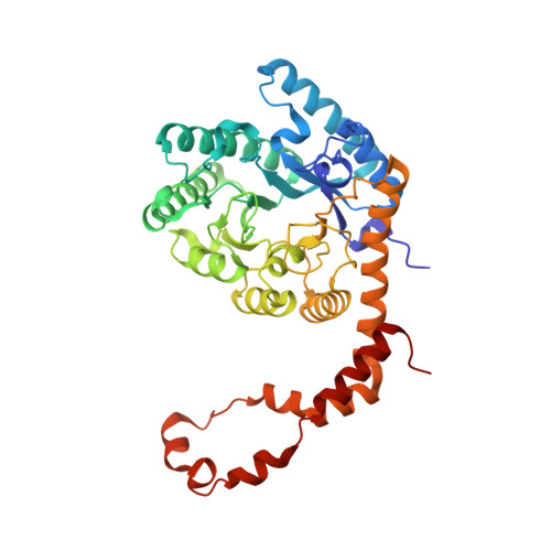
    We introduce a liquid application method for time-resolved analyses (LAMA), an in situ mixing approach for serial crystallography. Picoliter-sized droplets are shot onto chip-mounted protein crystals, achieving near-full ligand occupancy within theoretical diffusion times. We demonstrate proof-of-principle binding of GlcNac to lysozyme, and resolve glucose binding and subsequent ring opening in a time-resolved study of xylose isomerase.


  • Organizational Affiliation
    • Max-Planck-Institute for Structure and Dynamics of Matter, Department for Atomically Resolved Dynamics, Hamburg, Germany.

Macromolecule Content 

  • Total Structure Weight: 43.55 kDa 
  • Atom Count: 3,358 
  • Modeled Residue Count: 387 
  • Deposited Residue Count: 388 
  • Unique protein chains: 1

Macromolecules

Find similar proteins by:|  3D Structure
Entity ID: 1
MoleculeChains  Sequence LengthOrganismDetailsImage
Xylose isomerase388Streptomyces pseudogriseolusMutation(s): 0 
Gene Names: xylA
EC: 5.3.1.5
UniProt
Find proteins for P24300 (Streptomyces pseudogriseolus)
Explore P24300 
Go to UniProtKB:  P24300
Entity Groups
Sequence Clusters30% Identity50% Identity70% Identity90% Identity95% Identity100% Identity
UniProt GroupP24300
Sequence Annotations
Expand
Reference Sequence

Experimental Data & Validation

Experimental Data

  • Method: X-RAY DIFFRACTION
  • Resolution: 1.85 Å
  • R-Value Free:  0.196 (Depositor), 0.194 (DCC) 
  • R-Value Work:  0.165 (Depositor) 
  • R-Value Observed: 0.167 (Depositor) 
Space Group: I 2 2 2
Unit Cell:
Length ( Å )Angle ( ˚ )
a = 94.62α = 90
b = 99.38β = 90
c = 87.63γ = 90
Software Package:
Software NamePurpose
PHENIXrefinement
XDSdata reduction
XSCALEdata scaling
PHASERphasing

Structure Validation

View Full Validation Report


Ligand Structure Quality Assessment 


Entry History 

Deposition Data

Revision History  (Full details and data files)

  • Version 1.0: 2019-10-02
    Type: Initial release
  • Version 1.1: 2019-10-09
    Changes: Data collection, Database references
  • Version 1.2: 2019-11-20
    Changes: Derived calculations, Refinement description
  • Version 1.3: 2020-07-29
    Type: Remediation
    Reason: Carbohydrate remediation
    Changes: Data collection, Derived calculations, Structure summary
  • Version 1.4: 2024-05-15
    Changes: Data collection, Database references, Structure summary