Protein kinase B controls Mycobacterium tuberculosis growth via phosphorylation of the transcriptional regulator Lsr2 at threonine 112.
Alqaseer, K., Turapov, O., Barthe, P., Jagatia, H., De Visch, A., Roumestand, C., Wegrzyn, M., Bartek, I.L., Voskuil, M.I., O'Hare, H.M., Ajuh, P., Bottrill, A.R., Witney, A.A., Cohen-Gonsaud, M., Waddell, S.J., Mukamolova, G.V.(2019) Mol Microbiol 112: 1847-1862
- PubMed: 31562654 Search on PubMedSearch on PubMed Central
- DOI: https://doi.org/10.1111/mmi.14398
- Primary Citation Related Structures:
6QKP, 6QKQ - PubMed Abstract:
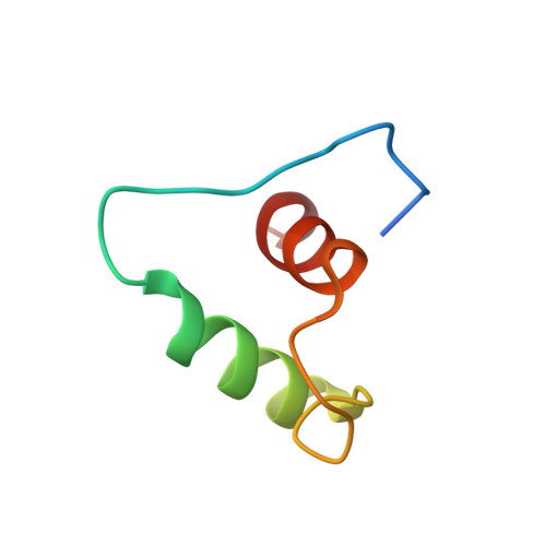
Mycobacterium tuberculosis (Mtb) is able to persist in the body through months of multi-drug therapy. Mycobacteria possess a wide range of regulatory proteins, including the protein kinase B (PknB) which controls peptidoglycan biosynthesis during growth. Here, we observed that depletion of PknB resulted in specific transcriptional changes that are likely caused by reduced phosphorylation of the H-NS-like regulator Lsr2 at threonine 112. The activity of PknB towards this phosphosite was confirmed with purified proteins, and this site was required for adaptation of Mtb to hypoxic conditions, and growth on solid media. Like H-NS, Lsr2 binds DNA in sequence-dependent and non-specific modes. PknB phosphorylation of Lsr2 reduced DNA binding, measured by fluorescence anisotropy and electrophoretic mobility shift assays, and our NMR structure of phosphomimetic T112D Lsr2 suggests that this may be due to increased dynamics of the DNA-binding domain. Conversely, the phosphoablative T112A Lsr2 had increased binding to certain DNA sites in ChIP-sequencing, and Mtb containing this variant showed transcriptional changes that correspond with the change in DNA binding. In summary, PknB controls Mtb growth and adaptations to the changing host environment by phosphorylating the global transcriptional regulator Lsr2.
- Leicester Tuberculosis Research Group, Department of Respiratory Sciences, University of Leicester, Leicester, LE2 9HN, UK.
Organizational Affiliation: