6QIX | pdb_00006qix

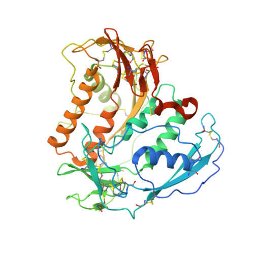
The crystal structure of Trichuris muris p43


Experimental Data Snapshot

  • Method: X-RAY DIFFRACTION
  • Resolution: 1.65 Å
  • R-Value Free: 
    0.189 (Depositor), 0.192 (DCC) 
  • R-Value Work: 
    0.150 (Depositor), 0.154 (DCC) 
  • R-Value Observed: 
    0.152 (Depositor) 

Starting Model: other
View more details

wwPDB Validation 3D Report Full Report

Validation slider image for 6QIX

Ligand Structure Quality Assessment 


This is version 1.3 of the entry. See complete history

Literature

The major secreted protein of the whipworm parasite tethers to matrix and inhibits interleukin-13 function.

Bancroft, A.J.Levy, C.W.Jowitt, T.A.Hayes, K.S.Thompson, S.Mckenzie, E.A.Ball, M.D.Dubaissi, E.France, A.P.Bellina, B.Sharpe, C.Mironov, A.Brown, S.L.Cook, P.C.S MacDonald, A.Thornton, D.J.Grencis, R.K.

(2019) Nat Commun 10: 2344-2344

  • DOI: https://doi.org/10.1038/s41467-019-09996-z
  • Primary Citation Related Structures: 
    6QIX

  • PubMed Abstract: 

    Infection by soil transmitted parasitic helminths, such as Trichuris spp, are ubiquitous in humans and animals but the mechanisms determining persistence of chronic infections are poorly understood. Here we show that p43, the single most abundant protein in T. muris excretions/secretions, is non-immunogenic during infection and has an unusual sequence and structure containing subdomain homology to thrombospondin type 1 and interleukin (IL)-13 receptor (R) α2. Binding of p43 to IL-13, the key effector cytokine responsible for T. muris expulsion, inhibits IL-13 function both in vitro and in vivo. Tethering of p43 to matrix proteoglycans presents a bound source of p43 to facilitate interaction with IL-13, which may underpin chronic intestinal infection. Our results suggest that exploiting the biology of p43 may open up new approaches to modulating IL-13 function and control of Trichuris infections.


  • Organizational Affiliation
    • Lydia Becker Institute for Immunology and Inflammation, Manchester, M13 9PT, UK. allison.j.bancroft@manchester.ac.uk.

Macromolecule Content 

  • Total Structure Weight: 92.96 kDa 
  • Atom Count: 6,509 
  • Modeled Residue Count: 694 
  • Deposited Residue Count: 788 
  • Unique protein chains: 1

Macromolecules

Find similar proteins by:|  3D Structure
Entity ID: 1
MoleculeChains  Sequence LengthOrganismDetailsImage
P43
A, B
394Trichuris murisMutation(s): 0 

Small Molecules

Ligands 5 Unique
IDChains Name / Formula / InChI Key2D Diagram3D Interactions
P6G

Query on P6G


Download:Ideal Coordinates CCD File
E [auth A],
F [auth A],
N [auth B],
O [auth B]
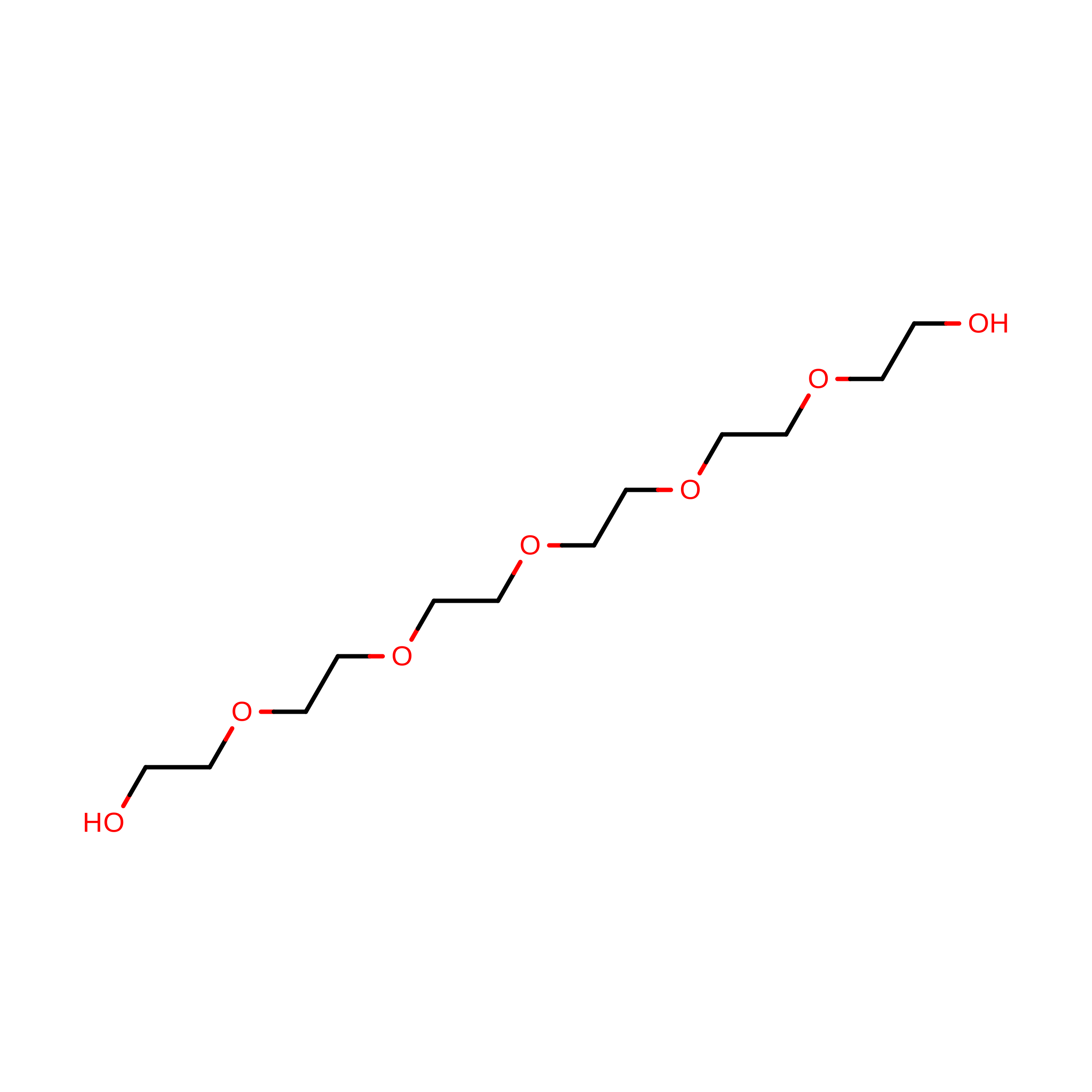
HEXAETHYLENE GLYCOL
C12 H26 O7
IIRDTKBZINWQAW-UHFFFAOYSA-N
NAG

Query on NAG


Download:Ideal Coordinates CCD File
C [auth A],
H [auth A],
M [auth B],
P [auth B]
2-acetamido-2-deoxy-beta-D-glucopyranose
C8 H15 N O6
OVRNDRQMDRJTHS-FMDGEEDCSA-N
PEG

Query on PEG


Download:Ideal Coordinates CCD File
J [auth A],
K [auth A],
Q [auth B],
R [auth B],
S [auth B]
DI(HYDROXYETHYL)ETHER
C4 H10 O3
MTHSVFCYNBDYFN-UHFFFAOYSA-N
ACT

Query on ACT


Download:Ideal Coordinates CCD File
D [auth A],
G [auth A],
L [auth A],
T [auth B]
ACETATE ION
C2 H3 O2
QTBSBXVTEAMEQO-UHFFFAOYSA-M
CA

Query on CA


Download:Ideal Coordinates CCD File
I [auth A]CALCIUM ION
Ca
BHPQYMZQTOCNFJ-UHFFFAOYSA-N

Experimental Data & Validation

Experimental Data

  • Method: X-RAY DIFFRACTION
  • Resolution: 1.65 Å
  • R-Value Free:  0.189 (Depositor), 0.192 (DCC) 
  • R-Value Work:  0.150 (Depositor), 0.154 (DCC) 
  • R-Value Observed: 0.152 (Depositor) 
Space Group: P 1 21 1
Unit Cell:
Length ( Å )Angle ( ˚ )
a = 47.682α = 90
b = 164.834β = 110.37
c = 61.169γ = 90
Software Package:
Software NamePurpose
PHENIXrefinement
DIALSdata reduction
DIALSdata scaling
PHASERphasing

Structure Validation

View Full Validation Report


Ligand Structure Quality Assessment 


Entry History 

& Funding Information

Deposition Data

  • Released Date: 2019-06-12 
  • Deposition Author(s): Levy, C.W.

Funding OrganizationLocationGrant Number
Wellcome TrustUnited KingdomWT100290MA
Wellcome TrustUnited KingdomZ10661/Z/18/Z

Revision History  (Full details and data files)

  • Version 1.0: 2019-06-12
    Type: Initial release
  • Version 1.1: 2020-07-29
    Type: Remediation
    Reason: Carbohydrate remediation
    Changes: Data collection, Derived calculations, Structure summary
  • Version 1.2: 2024-05-01
    Changes: Data collection, Database references, Refinement description, Structure summary
  • Version 1.3: 2024-10-23
    Changes: Structure summary