6QH3 | pdb_00006qh3

Catalytic domain of the human ubiquitin-conjugating enzyme UBE2S C118M


Experimental Data Snapshot

  • Method: X-RAY DIFFRACTION
  • Resolution: 2.90 Å
  • R-Value Free: 
    0.230 (Depositor), 0.217 (DCC) 
  • R-Value Work: 
    0.184 (Depositor) 
  • R-Value Observed: 
    0.187 (Depositor) 

Starting Model: experimental
View more details

wwPDB Validation 3D Report Full Report

Validation slider image for 6QH3

This is version 1.2 of the entry. See complete history

Literature

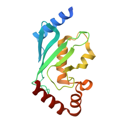
Autoinhibition Mechanism of the Ubiquitin-Conjugating Enzyme UBE2S by Autoubiquitination.

Liess, A.K.L.Kucerova, A.Schweimer, K.Yu, L.Roumeliotis, T.I.Diebold, M.Dybkov, O.Sotriffer, C.Urlaub, H.Choudhary, J.S.Mansfeld, J.Lorenz, S.

(2019) Structure 27: 1195-1210.e7

  • DOI: https://doi.org/10.1016/j.str.2019.05.008
  • Primary Citation Related Structures: 
    6QH3, 6QHK

  • PubMed Abstract: 

    Ubiquitin-conjugating enzymes (E2s) govern key aspects of ubiquitin signaling. Emerging evidence suggests that the activities of E2s are modulated by posttranslational modifications; the structural underpinnings, however, are largely unclear. Here, we unravel the structural basis and mechanistic consequences of a conserved autoubiquitination event near the catalytic center of E2s, using the human anaphase-promoting complex/cyclosome-associated UBE2S as a model system. Crystal structures we determined of the catalytic ubiquitin carrier protein domain combined with MD simulations reveal that the active-site region is malleable, which permits an adjacent ubiquitin acceptor site, Lys +5 , to be ubiquitinated intramolecularly. We demonstrate by NMR that the Lys +5 -linked ubiquitin inhibits UBE2S by obstructing its reloading with ubiquitin. By immunoprecipitation, quantitative mass spectrometry, and siRNA-and-rescue experiments we show that Lys +5 ubiquitination of UBE2S decreases during mitotic exit but does not influence proteasomal turnover of this E2. These findings suggest that UBE2S activity underlies inherent regulation during the cell cycle.


  • Organizational Affiliation
    • Rudolf Virchow Center for Experimental Biomedicine, University of Würzburg, 97080 Würzburg, Germany.

Macromolecule Content 

  • Total Structure Weight: 35.08 kDa 
  • Atom Count: 2,270 
  • Modeled Residue Count: 295 
  • Deposited Residue Count: 312 
  • Unique protein chains: 1

Macromolecules

Find similar proteins by:|  3D Structure
Entity ID: 1
MoleculeChains  Sequence LengthOrganismDetailsImage
Ubiquitin-conjugating enzyme E2 SA [auth B],
B [auth A]
156Homo sapiensMutation(s): 1 
Gene Names: UBE2SE2EPFOK/SW-cl.73
EC: 2.3.2.23
UniProt & NIH Common Fund Data Resources
Find proteins for Q16763 (Homo sapiens)
Explore Q16763 
Go to UniProtKB:  Q16763
PHAROS:  Q16763
GTEx:  ENSG00000108106 
Entity Groups
Sequence Clusters30% Identity50% Identity70% Identity90% Identity95% Identity100% Identity
UniProt GroupQ16763
Sequence Annotations
Expand
Reference Sequence

Experimental Data & Validation

Experimental Data

  • Method: X-RAY DIFFRACTION
  • Resolution: 2.90 Å
  • R-Value Free:  0.230 (Depositor), 0.217 (DCC) 
  • R-Value Work:  0.184 (Depositor) 
  • R-Value Observed: 0.187 (Depositor) 
Space Group: P 65
Unit Cell:
Length ( Å )Angle ( ˚ )
a = 84.619α = 90
b = 84.619β = 90
c = 87.832γ = 120
Software Package:
Software NamePurpose
PHENIXrefinement
XDSdata reduction
Aimlessdata scaling
PHASERphasing

Structure Validation

View Full Validation Report


Entry History 

& Funding Information

Deposition Data


Funding OrganizationLocationGrant Number
German Research FoundationGermanyEmmy Noether LO2003/1-1
German Research FoundationGRK 2243

Revision History  (Full details and data files)

  • Version 1.0: 2019-07-17
    Type: Initial release
  • Version 1.1: 2019-08-14
    Changes: Data collection, Database references
  • Version 1.2: 2024-01-24
    Changes: Data collection, Database references, Refinement description